
Introduction
- Group of genetically and clinically distinct disorders with few overlapping features.
- Most common phakomatoses.
- Subtypes:
- NF1 – Chromosome 17 – Neurofibromin (The word “Neurofibromastosis” has 17 letters!).
- NF2 – Chromosome 22 (2 is 22).
- NF3 – non-NF1/NF2 / Schwannomatosis / nonvestibular shwannomas.
- NF5- Segmental NF1 (old term).
Neurofibromatosis 1
- AKA von Recklinghausen disease.
- Autosomal dominant. Prevalence 1:3000.
Diagnostic criteria for NF-1
- Cutaneous Lesions
- • ≥6 café au lait spots(earliest manifestation). Prepubertal:≥0.5cm and Postpubertal: ≥ 1.5cm
- Freckling of armpits or groin.
- ≥ 2 neurofibromas(any type).
- Plexiform neurofibroma.
- Eye Abnormalities
- • ≥ 2 Lisch nodules(pigmented iris hamartomas).
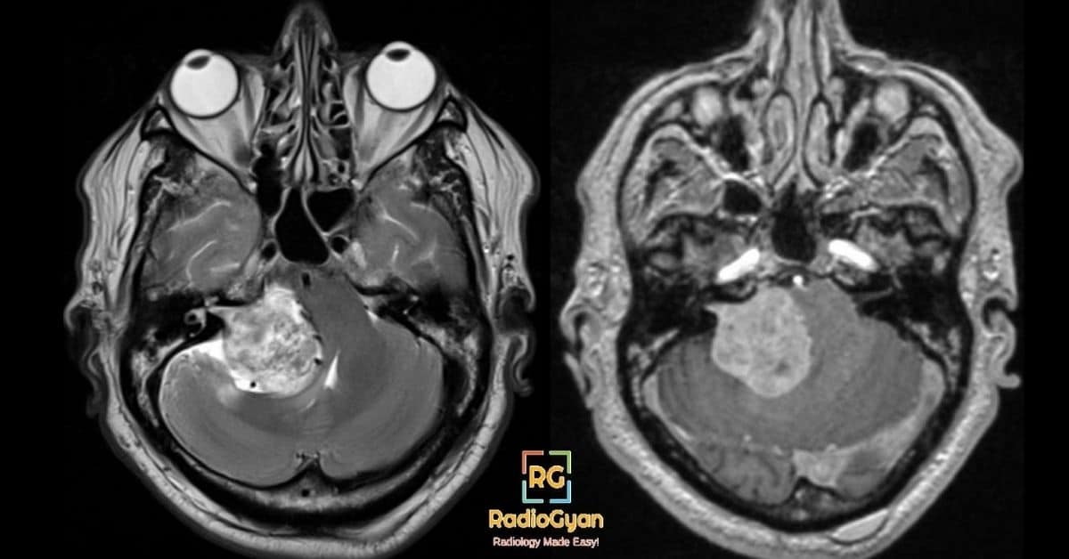
- Optic pathway pilocytic astrocytoma.
- Distinctive Bone Lesion
- Sphenoid dysplasia/absence
- Long bone cortex dysplasia/thinning
- Family History
- First-degree relative with NF1
- Neurofibromas:
- Arise commonly from superficial nerves.
- US: Fusiform and central. Nerve entering and exiting lesion.
- CT: Hypoattenuating with minimal enhancement.
- MRI: T2 hyperintense with central hypointensity – Target sign.
- Plexiform neurofibroma – Diffusely infiltrating. 10% malignant transformation.
- Others:
- Sphenoid wing dysplasia—hypoplastic/absent greater wing ± lesser wing giving rise to ‘bare orbit’ sign on plain film).
- Enlarged skull base foramina due to neurofibromas.
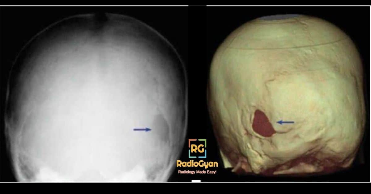
- Asterion defect : Lucent defects in the calvarium near the lambdoid sutures (refer image above).
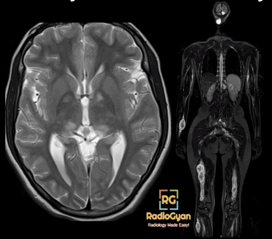
- Waxing and waning T2 hyperintense foci without mass effect involving white matter, basal ganglia, thalamus and cerebellum.
- Optic pathway glioma and other brain gliomas.
- Hydrocephalus.
- Arterial abnormalities.
- Kyphoscoliosis, especially of the thoracic spine.
- Dural ectasia.
- Spinal “dumb-bell” neurofibromas.
- Ribbon ribs.
- Abdominal and thoracic neurofibromas.
- Neuroendocrine tumors, paragangliomas GISTs and carcinoids.
- Anterolateral tibial bowing
- Focal gigantism
- Stenosis, aneurysms and AVMs.
Neurofibromatosis 2
Mnemonic for NF-2 – MISME
Multiple Inherited Schwannomas, Meningiomas and Ependymomas.
- Less common than NF1.
- Misnomer as not associated with neurofibromas.
Diagnostic criteria for NF-2
- Bilateral vestibular schwannomas.
- Any two of the following:
- (a) Unilateral vestibular schwannoma.
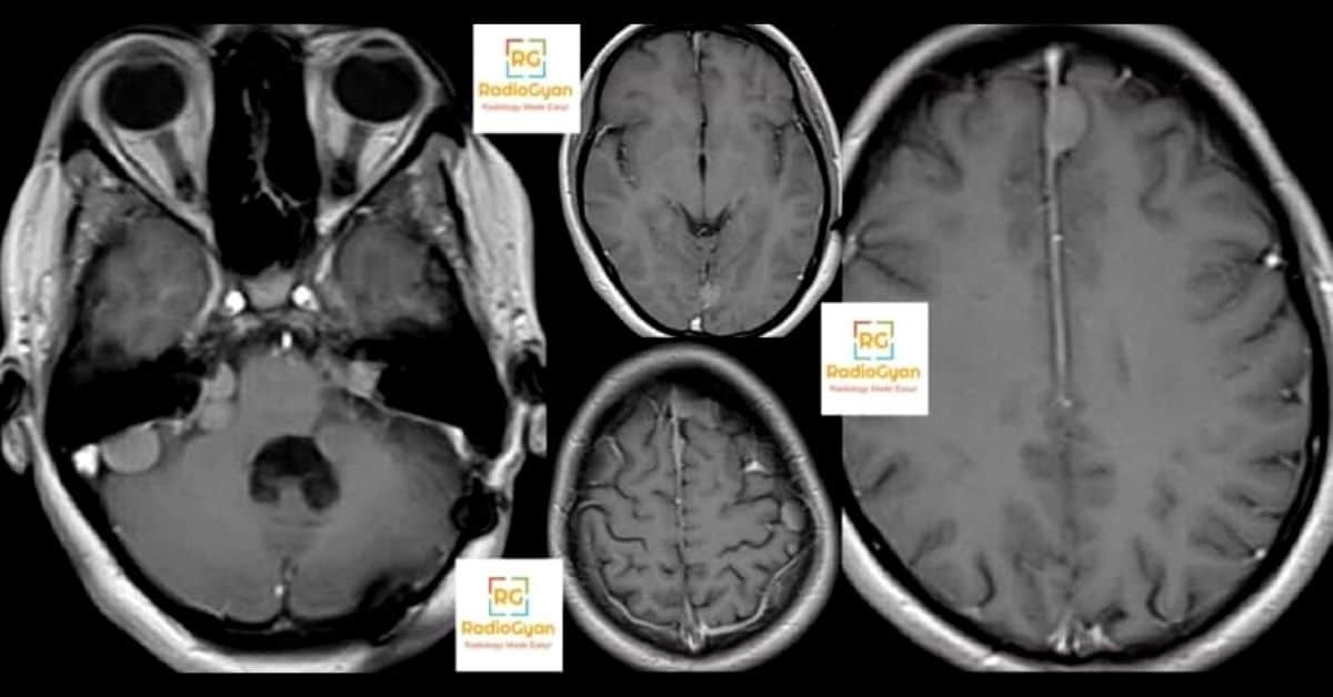
- (b) Any two of: meningioma, nonvestibular schwannoma, glioma (especially ependymoma), neurofibroma or juvenile cataract.
- (c) First-degree relative with NF2.
- ≥2 meningiomas + one of the following:
- (a) Unilateral vestibular schwannoma.
- (b) Any two of: nonvestibular schwannoma, glioma (especially ependymoma), neurofibroma or juvenile cataract.
Radiology signs associated with Neurofibromas
- Entering or exiting nerve sign: Lesion tapers on either end leading to the normal nerve.
- 🎯 Target sign: Seen on T2W images. Central T2 hypointensity due to central fibrocollagenous core and peripheral myxomatous T2 hyperintensity.
- Fascicular sign: Multiple ring-like structures, which appear as hypointense foci within the hyperintense area on T2W images, possibly reflecting the fascicular bundles.
- Split fat sign: Presence of fat at the upper and lower poles of a lesion on T1W images, which is suggestive of the intermuscular location of the lesion.
Neurofibroma vs Schwannoma?
Neurofibromas arise from the nerve while schwannomas arise from the nerve sheath, hence the former are central while the latter are eccenteric.
| Neurofibromas | Schwannomas | |
|---|---|---|
| Location | Central | Eccenteric |
| Target sign | More common | Less common |
| T2 hyperintense rim | Rare | Common |
| Intratumoral cysts | Rare | Common |
| Post-contrast enhancement | Central | Peripheral |
Signs of malignant peripheral nerve sheath tumors
Imaging features of Malignant Peripheral Nerve Sheath tumors
- Larger than 5 cm.
- Rapid increase in size.
- More heterogeneous. with necrosis and hemorrhage.
- Perilesional edema or infiltration.
- Bony destruction.
- Increased uptake on PET-CT.
References

Improve Content And Help Your Colleagues!
Found an error in the post? Please let us know using our contact page and we will update it with due credits!
If you wish to contribute radiological images for this case (or any other article on the website), you can submit them here and you will be duly credited: Submit radiology case
Sir you are best, great contribution for residents.
Thank you for your kind words, Asim :).
Many thanks, It is very simple and easy way to give information.
Glad you liked it Dr. Elshamsy.
Do check out more simplified radiology videos here: https://www.youtube.com/channel/UCrq_N7nwDk_Wo_AGhNakrMA?sub_confirmation=1