
Tuberculomas and gliomas are distinct brain pathologies, but they can share overlapping features on MRI, making differentiation challenging. Below is a detailed comparison of their radiological characteristics:
MRI Features: Tuberculomas vs. Gliomas
| Feature | Tuberculomas | Gliomas |
|---|---|---|
| Lesion Type | Granulomatous inflammation with caseous necrosis or non-caseating granulomas | Neoplastic growth; can be low-grade (benign) or high-grade (malignant) |
| T1-Weighted Imaging | Iso- to hypointense; caseating granulomas may show hyperintense rim | Hypointense to isointense; high-grade gliomas may show heterogeneous signal intensities |
| T2-Weighted Imaging | Hypointense (caseating granulomas) or hyperintense (non-caseating granulomas); surrounded by edema | Hyperintense with surrounding vasogenic edema; heterogeneous in higher grades |
| FLAIR Imaging | Partial suppression in caseating granulomas with liquefaction | Hyperintense signal; useful for delineating tumor margins and edema |
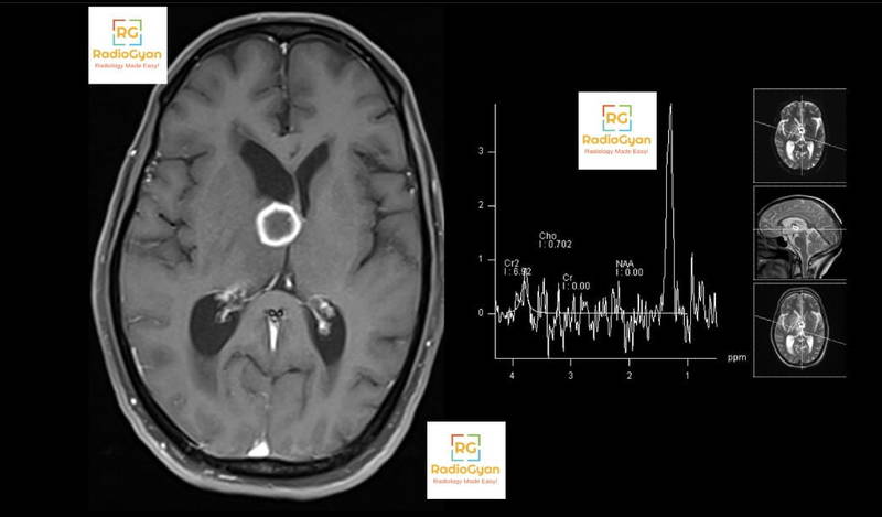
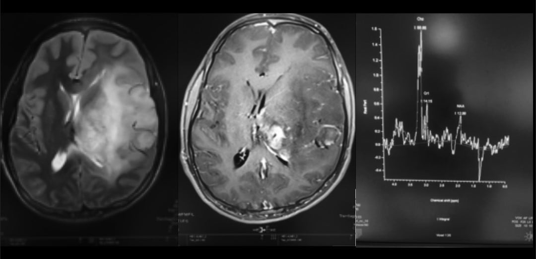
| Contrast Enhancement | Rim enhancement (caseating granulomas) or homogeneous enhancement (non-caseating granulomas) | Ring or nodular enhancement in high-grade gliomas; low-grade gliomas may lack enhancement |
| Diffusion-Weighted Imaging (DWI) | No restricted diffusion in most cases; variable in liquefactive necrosis | Restricted diffusion in areas of high cellularity, especially in high-grade gliomas |
| MR Spectroscopy | Elevated lipid-lactate peaks, reduced N-acetylaspartate (NAA)/creatine (Cr) ratio | Elevated choline (Cho), reduced NAA, and increased Cho/NAA ratio due to tumor metabolism |
| Edema and Mass Effect | Vasogenic edema surrounding lesions | Prominent vasogenic edema with significant mass effect in high-grade gliomas |
| Calcification | May show calcifications, especially in healed tuberculomas | Rarely calcified, except in oligodendrogliomas |
| Multiplicity | Often multiple lesions | Can be solitary or multifocal; multifocality suggests higher grade |
| Location | Predominantly basal ganglia, thalamus, or cortical gray matter | Can occur anywhere; infiltrative growth pattern common |
| Meningeal Involvement | May show meningeal enhancement | Rarely involves meninges unless there is leptomeningeal spread |
Key Differentiators
- Contrast Enhancement Patterns:
- Spectroscopy Findings:
- Edema Characteristics:
- Multiplicity and Distribution:
- Clinical Correlation:
- Tuberculomas are more common in endemic regions for tuberculosis or immunocompromised patients.
- Gliomas are associated with genetic mutations (e.g., IDH mutation) and are more frequent in older populations7.
Conclusion
While MRI features provide valuable clues, there is significant overlap between tuberculomas and gliomas. Advanced imaging techniques like MR spectroscopy, diffusion-weighted imaging, and clinical context are crucial for differentiation. A definitive diagnosis often requires histopathological confirmation through biopsy.
Citations:
- https://emedicine.medscape.com/article/344862-overview
- https://pmc.ncbi.nlm.nih.gov/articles/PMC8648135/
- https://onlinelibrary.wiley.com/doi/10.1155/2022/1955512
- https://www.frontiersin.org/journals/veterinary-science/articles/10.3389/fvets.2019.00286/full
- https://www.frontiersin.org/journals/radiology/articles/10.3389/fradi.2022.809373/full
- https://radiopaedia.org/articles/intracranial-tuberculous-granuloma
- https://ajronline.org/doi/10.2214/AJR.17.18457
- https://pmc.ncbi.nlm.nih.gov/articles/PMC3473894/
- https://pmc.ncbi.nlm.nih.gov/articles/PMC3686060/
- https://www.ncbi.nlm.nih.gov/pmc/articles/PMC11605145/
- https://pubmed.ncbi.nlm.nih.gov/38981890/
- https://www.semanticscholar.org/paper/0147f33e69f62c6c7de713676e9cc4ed825ff0f4
- https://pubmed.ncbi.nlm.nih.gov/39238702/
- https://www.semanticscholar.org/paper/61797785c50618aeff8d971c668f188af89c5260
- https://radiopaedia.org/articles/tuberculoma
- https://pmc.ncbi.nlm.nih.gov/articles/PMC6384409/
- https://onlinelibrary.wiley.com/doi/10.1155/2015/202806
- https://www.ncbi.nlm.nih.gov/pmc/articles/PMC10289839/
- https://www.ncbi.nlm.nih.gov/pmc/articles/PMC10810769/
- https://pubmed.ncbi.nlm.nih.gov/36576544/
- https://www.semanticscholar.org/paper/72bcc1dddcbca5c1d694d881cb5b47bd49f4a821
- https://www.ncbi.nlm.nih.gov/pmc/articles/PMC7817637/
- https://www.mayoclinic.org/diseases-conditions/glioma/symptoms-causes/syc-20350251
- https://www.youtube.com/watch?v=jvCRYT9RTFE
- https://www.hopkinsmedicine.org/health/conditions-and-diseases/gliomas
- https://pubs.rsna.org/doi/abs/10.1148/radiol.213063
- https://radiopaedia.org/cases/low-grade-glioma-1
- https://www.mdanderson.org/cancerwise/glioma-vs–glioblastoma–what-is-the-difference-in-these-brain-tumors-treatment-diagnosis.h00-159537378.html
- https://radiopaedia.org/articles/glioblastoma-idh-wildtype
- https://pubs.rsna.org/doi/abs/10.1148/radiology.174.2.2153310
- https://www.mdpi.com/2379-139X/7/2/21
- https://journals.sagepub.com/doi/full/10.1177/02841851211062083
- https://www.ajnr.org/content/ajnr/16/9/1903.full.pdf
- https://www.polradiol.com/Magnetic-resonance-imaging-spectrum-of-intracranial-tubercular-lesions-one-disease,102231,0,2.html
- https://pubs.rsna.org/doi/abs/10.1148/radiology.138.1.7455100
- https://journals.plos.org/plosone/article/file?type=printable&id=10.1371%2Fjournal.pone.0241974
- https://www.nature.com/articles/s41598-020-67080-9
- https://cco.amegroups.org/article/view/15820/html

Improve Content And Help Your Colleagues!
Found an error in the post? Please let us know using our contact page and we will update it with due credits!
If you wish to contribute radiological images for this case (or any other article on the website), you can submit them here and you will be duly credited: Submit radiology case