


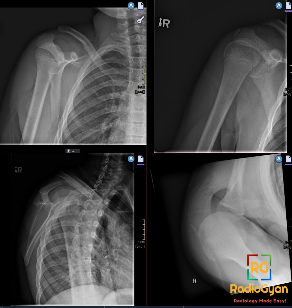
Young male presenting with right shoulder pain after a fall. What is your diagnosis?
Diagnosis and teaching points:
Diagnosis: Acromioclavicular joint injury. There is widening of the right AC joint with inferior displacement of the scapula.
Key facts for board exams:
- Apparent distal clavicular elevation is actually caused by inferior displacement of the scapula and upper extremity under gravity.
- Zanca view (10–15° cephalad tilt) optimally profiles the AC joint by eliminating overlying scapular spine shadow.
- The conoid and trapezoid ligaments provide primary vertical stability, whereas the acromioclavicular capsule resists horizontal translation.
- Axillary projection is mandatory when assessing high-grade injuries to rule out posterior clavicular displacement into the trapezius.
- Weighted stress views are no longer routinely recommended due to patient discomfort and inconsistent impact on clinical management.
Detailed teaching points:
- Clinical: Most common in young adult males during contact sports; presents with localized superior shoulder pain, deformity, and painful cross-body adduction.
- Etiology/Pathophys: Direct impact drives the acromion downward and medially while the clavicle remains fixed, leading to failure of the acromioclavicular joint capsule and adjacent deltotrapezial fascia.
- Radiograph: Normal joint space is under 5–7 mm; evaluation requires bilateral comparative views to detect asymmetric joint space widening and increased coracoclavicular distance.
- US: Real-time high-frequency imaging demonstrates capsular distention and ligament discontinuity, while dynamic external rotation unmasks occult horizontal joint instability.
- CT: Thin-section noncontrast 3D reformations identify associated coracoid fractures and define complex multiplanar osseous displacement for preoperative surgical planning.
- MRI: Fluid-sensitive fat-suppressed sequences demonstrate hyperintense ligament disruption and bone contusions, while evaluating for concurrent intra-articular SLAP tears or rotator cuff pathology.
- Signs: Piano-key sign – downward manual compression on the elevated clavicle reduces the deformity with immediate elastic rebound upon release.
- Frameworks: Rockwood classification: Type I (AC sprain), Type II (AC torn, CC sprained), Type III (AC/CC torn, 25–100% CC widening), Type IV (posterior displacement), Type V (>100% CC widening with fascia disruption), Type VI (subcoracoid).
- DDx: Distal clavicle fracture – focal cortical step-off with preserved coracoclavicular interval; coracoid base fracture – fractured coracoid process with intact ligaments; distal clavicular osteolysis – subchondral bone resorption and microcysts without acute traumatic vertical separation.
- Tx: Conservative sling immobilization and early rehabilitation for Rockwood Types I–II; surgical coracoclavicular reconstruction for Types IV–VI, failed conservative therapy, or high-demand overhead athletes.
OSCE Questions
Question: What classification system is most commonly used to grade this injury?
The Rockwood classification.
Question: Which specialized angled radiographic projection best evaluates this joint?
Zanca view (anteroposterior view with 10 to 15 degrees of cephalad angulation).
Question: What physical examination sign indicates vertical instability in this injury?
Piano-key sign.
Question: Disruption of which ligament complex causes vertical instability in this injury?
Coracoclavicular (CC) ligament complex (conoid and trapezoid ligaments).
Question: Which radiographic projection is essential to detect posterior displacement in this injury?
Axillary (or lateral) view, assessing for Rockwood Type IV injury.
MCQ Questions
1. Which radiographic view uses 10 to 15 degrees of cephalad beam angulation to project the acromioclavicular joint clear of overlapping osseous structures?
A. Standard anteroposterior view
B. Axillary lateral view
C. Zanca view
D. Scapular Y view
Answer: C. Zanca view. The Zanca view utilizes 10 to 15 degrees of cephalad beam angulation to optimize acromioclavicular joint visualization by reducing bony overlap from the acromion and scapular spine.
2. According to the Rockwood classification of acromioclavicular joint injuries, which subtype is defined by posterior displacement of the distal clavicle into or through the trapezius?
A. Type III
B. Type IV
C. Type V
D. Type VI
Answer: B. Type IV. Rockwood Type IV injuries are defined by complete acromioclavicular and coracoclavicular disruption with posterior displacement of the distal clavicle into or through the trapezius muscle.
3. Which radiographic finding specifically differentiates a Rockwood Type V acromioclavicular injury from a standard Rockwood Type III separation?
A. Increase in coracoclavicular distance exceeding 100 percent
B. Posterior displacement of the distal clavicle
C. Displacement of the clavicle inferior to the acromion
D. Widening of the joint space without vertical displacement
Answer: A. Increase in coracoclavicular distance exceeding 100 percent. Rockwood Type V represents severe vertical displacement characterized by deltotrapezial fascia disruption and coracoclavicular distance widening greater than 100 percent relative to the normal contralateral shoulder.
4. On shoulder radiographs, which imaging feature best differentiates a coracoid base fracture from isolated coracoclavicular ligament disruption in a patient with superior clavicular elevation?
A. Soft-tissue swelling over the superior shoulder
B. Widening of the acromioclavicular joint
C. Elevation of the distal clavicular shaft
D. Cortical disruption across the coracoid base
Answer: D. Cortical disruption across the coracoid base. Identification of a cortical fracture line through the coracoid base differentiates an osseous fracture from pure coracoclavicular ligamentous disruption despite similar superior clavicular displacement.
5. According to the Tossy classification for acromioclavicular joint injuries, which structural disruption pattern defines a Tossy Type II injury?
A. Sprain of acromioclavicular ligaments with intact joint
B. Acromioclavicular ligament rupture with coracoclavicular sprain
C. Complete rupture of acromioclavicular and coracoclavicular ligaments
D. Inferior subluxation of the clavicle beneath the coracoid
Answer: B. Acromioclavicular ligament rupture with coracoclavicular sprain. Tossy Type II is characterized by complete rupture of the acromioclavicular ligaments accompanied by a partial sprain of the coracoclavicular ligament complex.
Check out more such cases:

Improve Content And Help Your Colleagues!
Found an error in the post? Please let us know using our contact page and we will update it with due credits!
If you wish to contribute radiological images for this case (or any other article on the website), you can submit them here and you will be duly credited: Submit radiology case