


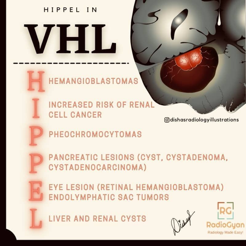
Von Hippel-Lindau (VHL) syndrome is an autosomal dominant, multi-system tumour-predisposition disorder caused by a germline variant in the VHL tumour-suppressor gene on chromosome 3p25.3. Loss of functional VHL protein leaves hypoxia-inducible factor 2-alpha (HIF-2α) unchecked, driving over-expression of VEGF and erythropoietin and a crop of characteristically hypervascular tumours and cysts across the CNS, retina, kidneys, adrenals, pancreas and inner ear. Radiologists are central to VHL care because most lesions are found, followed and treated on imaging long before they become symptomatic. This page keeps the classic HIPPEL mnemonic and adds the diagnostic criteria, tumour spectrum, imaging features, surveillance schedule and current treatment.
HIPPEL mnemonic for VHL syndrome
H – Hemangioblastomas (cerebellum, brainstem, spinal cord)
I – Increased risk of renal cell carcinoma (clear cell)
P – Pheochromocytomas (and paragangliomas)
P – Pancreatic lesions (cysts, serous cystadenoma, neuroendocrine tumours)
E – Eye (retinal hemangioblastoma) and Endolymphatic sac tumours
L – Liver and renal cysts (plus epididymal / broad-ligament cystadenomas)
What is VHL syndrome?
VHL is inherited in an autosomal dominant fashion with high penetrance, although roughly 20% of cases arise from a de novo mutation with no family history. The birth incidence is about 1 in 27,000 to 36,000. Because a working copy of the gene must be lost within a cell before a tumour develops (the two-hit model), lesions appear at different ages in different organs, and new lesions continue to arise throughout life — which is why lifelong surveillance, rather than a single work-up, defines management.
Tumours associated with VHL syndrome
| Organ / system | Characteristic lesion | Approx. lifetime prevalence | Key imaging features |
|---|---|---|---|
| Cerebellum, brainstem, spinal cord | Hemangioblastoma | 60–80% | Cyst with an avidly enhancing mural nodule; flow voids; frequently multiple |
| Retina | Retinal hemangioblastoma (capillary angioma) | ~50–60% | Enhancing retinal nodule with dilated feeding and draining vessels; often the earliest manifestation |
| Kidney | Clear cell renal cell carcinoma and cysts | RCC up to ~70% | Multiple, bilateral, often multifocal cystic and solid enhancing masses |
| Adrenal / sympathetic chain | Pheochromocytoma / paraganglioma | ~10–20% (higher in type 2) | Markedly T2-hyperintense, avidly enhancing mass; DOTATATE / MIBG avid |
| Pancreas | Serous cystadenoma, neuroendocrine tumour, true cysts | Cysts up to ~70% | Multiple cysts, microcystic serous cystadenoma, hypervascular NET |
| Inner ear (endolymphatic sac) | Endolymphatic sac tumour (ELST) | ~4–16% | Retrolabyrinthine petrous mass, intratumoral calcification, peripheral T1-hyperintense foci |
| Epididymis / broad ligament | Papillary cystadenoma | Up to ~60% of males (epididymal) | Cystic or solid paratesticular / adnexal lesion; usually benign |
Diagnostic criteria
A clinical diagnosis of VHL can be made on the pattern of tumours alone, and is confirmed by genetic testing:
- With a family history of VHL: a single characteristic tumour is sufficient — a CNS or retinal hemangioblastoma, clear cell RCC, pheochromocytoma, pancreatic neuroendocrine tumour or endolymphatic sac tumour.
- Without a family history: two or more characteristic tumours are required — typically two hemangioblastomas, or one hemangioblastoma plus a visceral tumour.
- Molecular confirmation: identifying a pathogenic or likely pathogenic germline VHL variant establishes the diagnosis even when clinical criteria are not yet met, and guides cascade testing of relatives.
VHL clinical subtypes
VHL is divided by pheochromocytoma risk, which correlates loosely with the type of germline variant. This matters for surveillance and for anaesthetic planning before any intervention.
| Subtype | Pheochromocytoma risk | RCC risk | Typical variant |
|---|---|---|---|
| Type 1 | Low | High | Deletions / truncating (loss of function) |
| Type 2A | High | Low | Missense |
| Type 2B | High | High | Missense |
| Type 2C | High (pheochromocytoma only) | Low / absent | Missense |
Imaging features by organ
- CNS hemangioblastoma: the commonest CNS tumour in VHL. Classically a cyst with an avidly enhancing mural nodule abutting a pial surface, with intratumoral or peritumoral flow voids. The cerebellum is the favourite site, followed by the spinal cord and brainstem. Multiplicity in a young patient should always prompt a search for VHL.
- Retinal hemangioblastoma: often the first lesion to appear, sometimes in childhood. Seen on fundoscopy and MRI as a small enhancing retinal nodule with prominent feeding and draining vessels; untreated lesions risk exudation, retinal detachment and blindness.
- Renal cell carcinoma and cysts: VHL RCC is clear cell type, tends to be multiple, bilateral and to recur, and is the leading cause of VHL-related death. Solid or solid-and-cystic enhancing masses are followed by size; the 3 cm threshold traditionally triggers nephron-sparing surgery to balance cancer control against preserving renal function.
- Pheochromocytoma: may be bilateral or extra-adrenal. Markedly T2-hyperintense and avidly enhancing, and functionally imaged with 68Ga-DOTATATE PET or MIBG. Biochemical screening should always precede any adrenal or renal surgery.
- Pancreatic lesions: a spectrum of true cysts, microcystic serous cystadenoma and hypervascular neuroendocrine tumours. Multiple pancreatic cysts in a young patient are a useful clue to the diagnosis; NETs are followed by size and are the lesions most likely to need resection or systemic therapy.
- Endolymphatic sac tumour (ELST): a locally aggressive papillary tumour of the posterior petrous temporal bone. CT shows a retrolabyrinthine lytic mass with intratumoral spiculated calcification; MRI shows peripheral T1-hyperintense foci. A cause of sensorineural hearing loss and vertigo that is easy to miss if the temporal bone is not scrutinised.
Surveillance schedule
Surveillance for affected and at-risk individuals is coordinated by multidisciplinary teams and starts in childhood. The schedule below reflects the international consensus and VHL Alliance surveillance guidelines; exact ages and intervals are individualised.
| Test | Begin at age | Interval | Target lesion |
|---|---|---|---|
| Dilated retinal (ophthalmological) exam | Infancy / diagnosis | Annually | Retinal hemangioblastoma |
| Blood pressure plus plasma or urine fractionated metanephrines | 5 years | Annually | Pheochromocytoma |
| Contrast-enhanced MRI brain and whole spine | 11 years | Every 2 years | CNS hemangioblastoma |
| Audiology (with internal auditory canal MRI if symptomatic) | 11 years | Every 2–3 years | Endolymphatic sac tumour |
| Abdominal MRI (kidneys, pancreas, adrenals) | 15 years | Annually / as indicated | RCC, pancreatic tumours, pheochromocytoma |
MRI is preferred over CT wherever possible to limit cumulative radiation across a lifetime of imaging, and whole-body MRI is being studied as a single-visit alternative. Ultrasound is a reasonable adjunct for renal follow-up but does not replace cross-sectional surveillance.
Treatment
The backbone of care remains surveillance with organ-sparing surgery when a lesion grows, becomes symptomatic or crosses a size threshold — for example nephron-sparing resection for renal tumours and resection of enlarging or symptomatic hemangioblastomas. Retinal lesions are treated with laser photocoagulation or cryotherapy, and pheochromocytomas are resected after appropriate alpha-blockade.
Belzutifan (Welireg), a first-in-class oral HIF-2α inhibitor, was approved by the FDA on 13 August 2021 for adults with VHL-associated RCC, CNS hemangioblastoma or pancreatic neuroendocrine tumour that does not require immediate surgery. In the pivotal LITESPARK-004 trial the renal cell carcinoma objective response rate was about 49%, with durable responses and meaningful shrinkage of hemangioblastomas and pancreatic lesions maintained at 50-month follow-up; anaemia and fatigue are the dominant toxicities. Belzutifan offers a systemic option that can defer or reduce repeated surgeries, and is reshaping how radiologists report and follow VHL tumour burden. Advanced or metastatic RCC not suited to belzutifan is treated with VEGF-directed agents such as pazopanib.
Frequently asked questions
References
- Ganeshan D, Menias CO, Pickhardt PJ, et al. Tumors in von Hippel-Lindau Syndrome: From Head to Toe—Comprehensive State-of-the-Art Review. RadioGraphics. 2018;38(3):849-866. PMID: 29601266.
- Leung RS, Biswas SV, Duncan M, Rankin S. Imaging features of von Hippel-Lindau disease. RadioGraphics. 2008;28(1):65-79. PMID: 18203931.
- Binderup MLM, Smerdel M, Borgwadt L, et al. von Hippel-Lindau disease: Updated guideline for diagnosis and surveillance. Eur J Med Genet. 2022;65(8):104538. PMID: 35709961.
- Daniels AB, Tirosh A, Huntoon K, et al. Guidelines for surveillance of patients with von Hippel-Lindau disease: Consensus statement of the International VHL Surveillance Guidelines Consortium and VHL Alliance. Cancer. 2023;129(19):2927-2940. PMID: 37337409.
- Jonasch E, Donskov F, Iliopoulos O, et al. Belzutifan for Renal Cell Carcinoma in von Hippel-Lindau Disease. N Engl J Med. 2021;385(22):2036-2046. PMID: 34818478.
- Srinivasan R, Iliopoulos O, Beckermann KE, et al. Belzutifan for von Hippel-Lindau disease-associated renal cell carcinoma and other neoplasms (LITESPARK-004): 50 months follow-up from a single-arm, phase 2 study. Lancet Oncol. 2025;26(5):571-582. PMID: 40228516.
Browse more high-yield revision aids on our radiology mnemonics collection.
Check out more visual mnemonics here:
Causes Of Micronodular Spleen – Visual Mnemonics

Pachymeningeal Thickening – Visual Mnemonics

Causes Of Patternless Bowel – Visual Mnemonics

Improve Content And Help Your Colleagues!
Found an error in the post? Please let us know using our contact page and we will update it with due credits!
If you wish to contribute radiological images for this case (or any other article on the website), you can submit them here and you will be duly credited: Submit radiology case