


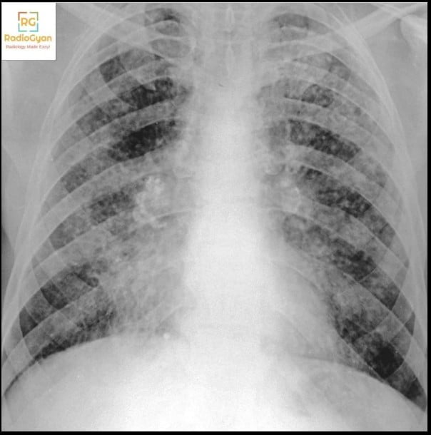
Apical fibrosis, also reported as upper lobe fibrosis, is scarring at the lung apex with volume loss, reticulation, traction bronchiectasis and architectural distortion that pulls the hilum and the trachea upwards. Previous tuberculosis and previous radiotherapy account for most cases, followed by sarcoidosis, ankylosing spondylitis, silicosis and chronic hypersensitivity pneumonitis. Thin biapical scarring on a chest radiograph is usually an incidental post-inflammatory finding.

Remember THE SCARS for the causes of apical lung fibrosis:
T – Tuberculosis
H – Histoplasmosis and other chronic granulomatous infection
E – Extrinsic allergic alveolitis (hypersensitivity pneumonitis)
S – Silicosis
C – Coal workers’ pneumoconiosis
A – Ankylosing spondylitis
R – Radiation
S – Sarcoidosis
Key facts for practice and radiology board exams
- Healed tuberculosis is the commonest cause worldwide. Post-primary disease favours the apical and posterior segments of the upper lobes and the superior segments of the lower lobes [1]. Post-tuberculosis lung disease now has a formal case definition: prior tuberculosis plus abnormality in at least two of lung function, symptoms and imaging [3].
- Laterality triages it faster than anything else. Unilateral means previous localised infection, radiotherapy, surgery or a superior sulcus tumour. Biapical and symmetrical means sarcoidosis, ankylosing spondylitis, pneumoconiosis or pleuroparenchymal fibroelastosis.
- Apical scarring is usually age, not disease. On CT in apparently healthy adults it was present in 5.5 percent overall: 1.6 percent at ages 20 to 39, 3.9 percent at 40 to 59 and 8.1 percent at 60 and over. Only 12.7 percent progressed over about four years [9].
- Ankylosing spondylitis: 6.9 percent of 303 patients in a systematic review had upper lobe fibrosis on HRCT, and it was the only abnormality that rose significantly with disease duration [5]. Check the spine and sacroiliac joints on the same study.
- Sarcoidosis: up to 40 percent progress to stage IV fibrotic disease with hilar retraction and fibrocystic change [6].
- Radiation fibrosis is usually confined to the treatment port, but conformal and stereotactic techniques produce atypical, focal patterns that no longer follow a port outline [7].
- A new mass or air crescent inside old apical fibrosis is a fungal ball until proven otherwise. A global burden model estimated that 21 to 35 percent of treated pulmonary tuberculosis leaves cavities, and about 22 percent of those develop chronic pulmonary aspergillosis [4], a modelled figure rather than a measured rate.
Causes of apical lung fibrosis and how to tell them apart
The eight causes are not equally likely, and most separate on the film itself. Work through laterality, then the company the fibrosis keeps.
| Cause | Typical pattern | What separates it from the rest |
|---|---|---|
| Previous tuberculosis | Asymmetrical upper zone fibrocavitary change with calcified granulomas and nodes, upward hilar retraction and tracheal shift | Calcification and residual cavities. A new mass or air crescent inside an old cavity is a mycetoma, not reactivation |
| Radiotherapy | Dense fibrosis with straight, non-anatomical margins following the old treatment port, with volume loss and traction bronchiectasis | Margins that cross fissures and ignore segmental anatomy. Conformal and stereotactic treatment give focal mass-like fibrosis instead |
| Sarcoidosis | Upper and mid zone perilymphatic nodularity progressing to conglomerate perihilar masses and marked upward hilar retraction | Nodules along fissures and bronchovascular bundles, plus symmetrical hilar nodes, often calcified |
| Ankylosing spondylitis | Bilateral apical fibrobullous disease with cysts and bullae, often colonised by a mycetoma | The spine: syndesmophytes, squared vertebrae and fused sacroiliac joints on the same films |
| Silicosis and coal workers’ pneumoconiosis | Upper zone nodules coalescing into progressive massive fibrosis, with peripheral emphysema | Eggshell calcification of hilar nodes, an occupational history, and masses that migrate hilarwards over time |
| Chronic fibrotic hypersensitivity pneumonitis | Reticulation and traction bronchiectasis on a background of mosaic attenuation and air trapping | The mosaic background and air trapping, not the zone. Its place in this mnemonic is the weakest: one HRCT series found the fibrosis was mid zone or non zonal with the apices and bases relatively spared [8] |
| Chronic granulomatous infection other than tuberculosis | Upper zone nodules, cavities and fibrosis indistinguishable from tuberculosis | Geography and host: histoplasmosis, and non-tuberculous mycobacteria in bronchiectatic or emphysematous lungs |
| Pleuroparenchymal fibroelastosis | Dense upper zone subpleural fibrosis and pleural thickening with severe upper lobe volume loss | A flattened chest wall, low body mass index and spared lower lobes |
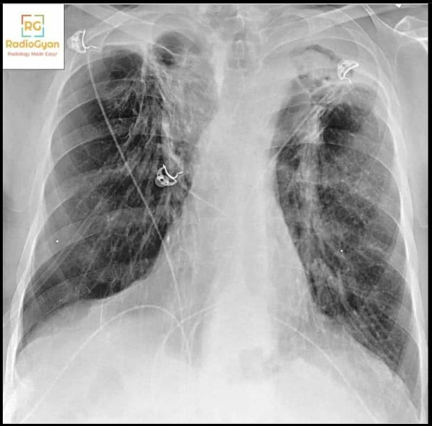
Apical scarring, apical cap and apical fibrosis are not the same thing
These three phrases turn up in the same reports and are not interchangeable, and only one of them needs a cause.
| Term | What it is | Significance |
|---|---|---|
| Apical scarring, biapical scarring | A descriptive radiograph term for thin linear or reticular opacity at one or both apices with minor volume loss | Usually incidental and post-inflammatory. No action if thin, smooth and unchanged |
| Pleuroparenchymal apical cap | A subpleural band of fibroelastotic tissue over the apex, becoming more common with age and with a lower body mass index [9] | Benign when stable. It is progression over serial imaging, not thickness, that separates a cap from pleuroparenchymal fibroelastosis [10] |
| Apical lung fibrosis, upper lobe fibrosis | Established parenchymal fibrosis with reticulation, traction bronchiectasis, architectural distortion and volume loss | Has a cause worth naming. Work through the differential below |
Why the apex
Gravity gives the upright apex the lowest blood flow and so the highest local oxygen tension, the textbook explanation for why tuberculosis, chronic histoplasmosis and progressive massive fibrosis all favour the upper zones. Goodwin and Des Prez, examining all three together, argued the better explanation is mechanical rather than metabolic: low apical perfusion also means sluggish lymph flow, so inhaled organisms, dust and antigen are cleared more slowly and the inflammatory response is prolonged [2]. The practical consequence is the same either way. Anything inhaled, poorly cleared and slow to resolve scars at the top.
Pleuroparenchymal fibroelastosis: the cause the mnemonic misses
Pleuroparenchymal fibroelastosis is upper lobe subpleural fibrosis with pleural thickening, marked upper lobe volume loss and relatively spared lower lobes, and it is in none of the mnemonics. It is the one entity here defined by change over time. The clinical criteria require bilateral subpleural dense consolidation in the upper lobes, documented radiologic progression, and exclusion of other causes, and the progression requirement exists specifically to exclude a pulmonary apical cap, which mimics it both radiologically and pathologically [10]. Patients are characteristically thin. The practical point: a single CT cannot make this diagnosis, and a comparison study can.
How to approach apical fibrosis on a chest radiograph or CT
- Compare with the oldest film you can find. Stability over years is more reassuring than any morphological argument.
- Decide unilateral or biapical first, then look for the company it keeps: calcified nodes, eggshell calcification, perilymphatic nodules, a fused spine, a clip line or a port edge.
- Look for a cavity, and look inside it. A soft tissue mass that moves on prone imaging is a mycetoma. New consolidation, a new cavity or new nodules in old fibrosis raise reactivation.
- Treat an asymmetric, nodular or enlarging cap as a lesion, not a cap. Contrast CT, and ask about shoulder or arm pain, Horner syndrome and rib destruction.
Related resources on RadioGyan
- CARPET mnemonic for apical lung diseases, the wider list that includes apical consolidation and cysts.
- Radiology signs index, including the Monod and air crescent signs of a mycetoma in a fibrotic cavity.
Frequently asked questions
References
- McAdams HP, Erasmus J, Winter JA. Radiologic manifestations of pulmonary tuberculosis. Radiol Clin North Am. 1995;33(4):655-678. PMID: 7610237.
- Goodwin RA, Des Prez RM. Apical localization of pulmonary tuberculosis, chronic pulmonary histoplasmosis, and progressive massive fibrosis of the lung. Chest. 1983;83(5):801-805. PMID: 6839825.
- Bisson GP, Allwood B, Byrne A, et al. Post-tuberculosis lung disease: a case definition for use in research studies. Lancet Infect Dis. 2026;26(7):e315-e325. PMID: 41232546.
- Denning DW, Pleuvry A, Cole DC. Global burden of chronic pulmonary aspergillosis as a sequel to pulmonary tuberculosis. Bull World Health Organ. 2011;89(12):864-872. PMID: 22271943.
- El Maghraoui A, Dehhaoui M. Prevalence and characteristics of lung involvement on high resolution computed tomography in patients with ankylosing spondylitis: a systematic review. Pulm Med. 2012;2012:965956. PMID: 22536503.
- Belperio JA, Fishbein MC, Abtin F, Channick J, Balasubramanian SA, Lynch JP 3rd. Pulmonary sarcoidosis: a comprehensive review: past to present. J Autoimmun. 2024;149:103107. PMID: 37865579.
- Ghaye B, Wanet M, El Hajjam M. Imaging after radiation therapy of thoracic tumors. Diagn Interv Imaging. 2016;97(10):1037-1052. PMID: 27567554.
- Adler BD, Padley SP, Muller NL, Remy-Jardin M, Remy J. Chronic hypersensitivity pneumonitis: high-resolution CT and radiographic features in 16 patients. Radiology. 1992;185(1):91-95. PMID: 1523340.
- Fukada A, Hozumi H, Fukuda T, et al. Clinical characteristics and prevalence of apical scarring at chest CT in a healthy population. Radiol Cardiothorac Imaging. 2026;8(1):e250190. PMID: 41711551.
- Enomoto Y, Nakamura Y, Satake Y, et al. Clinical diagnosis of idiopathic pleuroparenchymal fibroelastosis: a retrospective multicenter study. Respir Med. 2017;133:1-5. PMID: 29173442.
Check out more visual mnemonics here:

Intramedullary Lesions – Visual Mnemonics

Apple Core Sign Differentials – Visual Mnemonics
Ankle Tendons Anatomy- Visual Mnemonics

Improve Content And Help Your Colleagues!
Found an error in the post? Please let us know using our contact page and we will update it with due credits!
If you wish to contribute radiological images for this case (or any other article on the website), you can submit them here and you will be duly credited: Submit radiology case