
Bone age assessment plays a crucial role in pediatric radiology, helping to determine a child’s level of maturation and growth potential. X-ray imaging is commonly used to calculate bone age, providing valuable information for diagnosing various skeletal disorders, monitoring growth patterns, and assessing overall development. In this article, we will delve into the process of calculating bone age using X-rays, highlighting key considerations and steps involved.
What is Bone Age?
Bone age refers to the degree of skeletal maturation in relation to chronological age. It is determined by assessing the ossification centers and the fusion of growth plates in different bones, especially in the hand and wrist regions. The assessment compares the observed skeletal development with a standard reference set, typically represented by the Greulich-Pyle (GP) atlas or the Tanner-Whitehouse (TW) method.
Common conditions where pediatric bone age estimation is necessary include constitutional growth delay, precocious puberty, growth hormone deficiency, Turner syndrome, Down syndrome, and skeletal dysplasias.
The Process of Calculating Bone Age Using X-rays
- Patient Preparation: Before proceeding with bone age assessment, ensure that the patient is properly positioned and relaxed. It is essential to obtain high-quality X-ray images, so it may be necessary to stabilize the patient’s hand and wrist to reduce motion artifacts.
- Imaging Technique: The standard imaging technique for bone age assessment involves obtaining a posterior-anterior (PA) radiograph of the left hand and wrist. This projection allows for consistent visualization of the carpal bones, phalanges, and epiphysis.
- Image Analysis: Once the X-ray image is obtained, carefully analyze the radiograph to identify the relevant anatomical structures. Key landmarks to consider include:
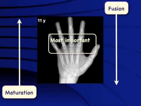
- Epiphyseal plates: Evaluate the presence, size, and degree of fusion of the epiphyseal plates in both the carpal bones and phalanges.
- Ossification centers: Observe the ossification centers in each bone, noting their appearance and development stage.
- Reference Atlas: Use a standard reference atlas such as the Greulich-Pyle (GP) or Tanner-Whitehouse (TW) atlas to compare the observed skeletal development with established reference values. These atlases consist of radiographic images corresponding to known ages, enabling estimation of bone age by matching similar patterns of skeletal maturity.
- The GP atlas was developed by Greulich and Pyle in the 1950s and is based on X-ray images of the left hand and wrist. It provides a set of standards for each age group, allowing doctors to compare an individual’s X-ray to the standard images and determine their bone age.
- The TW atlas, also known as the Tanner-Whitehouse-III (TW3) method, was developed later in the 1970s. It takes into account additional skeletal features along with the hand and wrist bones to estimate bone age. The TW3 method is considered more accurate than the GP method, especially in older children.
- Interpretation: Compare the identified ossification centers and epiphyseal plates in the patient’s X-ray image with the corresponding reference images in the atlas. Based on similarity, estimate the bone age by determining the closest match in terms of skeletal maturation.
- Reporting: Document your findings accurately and comprehensively in a radiology report. Include details about the patient, examination technique, key observations, estimated bone age, and any additional relevant information.
Pediatric Bone Age Calculators – Machine learning and AI based
Best online tools currently available for bone age calculation:
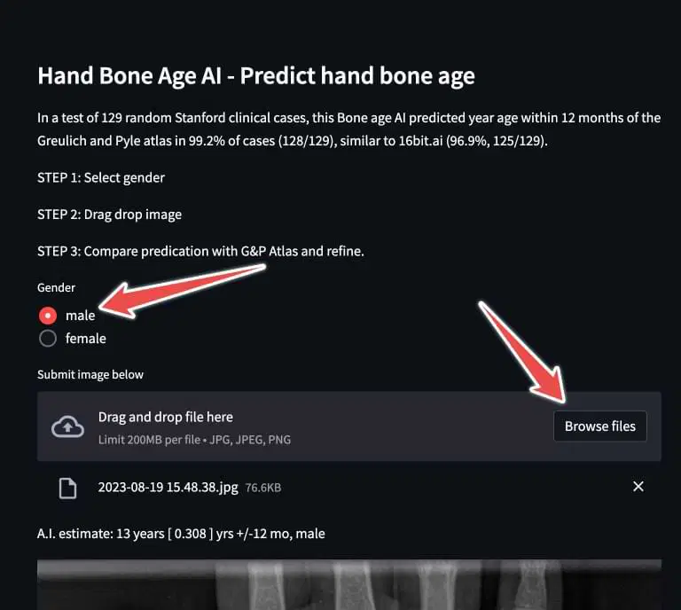
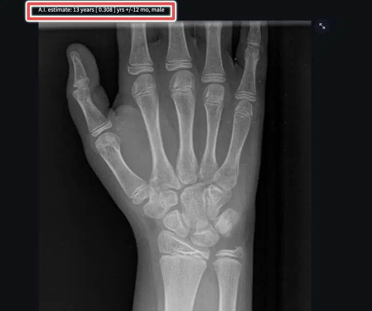
- Stanford Radiology Hand Bone Age AI – Automated
- FreeBoneAge.com – Automated
- 16bit.AI Bone Age Calculator – Paid
Example using Stanford tool
KerbyRadRes earlier used to provide a nice calculator for bone. This is also the first result that shows up on Google search. However the reference radiograph images have been removed. You can use the reporting template there, but without the images, it is not much of value.
Standard Atlas for Reference to Calculate Bone Age
These websites have normal radiographs for reference. Compare the radiographs you are evaluating with the xrays in the atlas to get an estimate of the bone age.
Bone Age Estimation Video Tutorial
Limitations and Considerations
It is important to note that bone age assessment using X-rays has its limitations and should be interpreted with caution. Some considerations include:
- Variability: There can be significant individual variation in bone age development, influenced by genetic factors, nutrition, and overall health.
- Ethnicity and Gender: Different populations and genders may have distinct patterns of skeletal maturation, necessitating appropriate reference standards for accurate interpretation.
- Developmental Disorders: Certain conditions, such as endocrine disorders or growth hormone deficiencies, can impact bone age development, requiring additional clinical evaluation.
- Radiation Exposure: As with any medical imaging procedure involving X-rays, practitioners should prioritize minimizing radiation exposure while maintaining diagnostic image quality.
Conclusion
Calculating bone age using X-rays is a valuable tool in pediatric radiology for assessing skeletal maturation and growth potential. By following a systematic approach, including patient preparation, proper imaging technique, image analysis, reference atlas comparison, interpretation, and reporting, accurate estimations can be made. However, it is crucial to consider the limitations and individual variations associated with bone age assessment when interpreting results.

Improve Content And Help Your Colleagues!
Found an error in the post? Please let us know using our contact page and we will update it with due credits!
If you wish to contribute radiological images for this case (or any other article on the website), you can submit them here and you will be duly credited: Submit radiology case
Corrected Version (Recommended):
Dear Sir,
Could you please grant us free full access to the AI-based bone age calculation tool?
Thank you very much for your kind support for a resource-limited country.
Best regards,
Songngin
Hey. I do not have access to the paid tool. The Stanford tool is free: http://www.xrayhead.com:8080/
xray result for a boy
A.I. estimate: 16 years [ 0.756 ] yrs +/-12 mo, male
how much he will be tall
Unfortunately we will not be able to help with that. Please talk to your physician.