


The axial, coronal and sagittal planes are the three cardinal planes used to describe position in the body and to display cross-sectional images. The quickest way to tell them apart is by what each one divides the body into:
- Axial (transverse) plane is horizontal, and divides the body into upper and lower. This is the plane CT data are acquired in.
- Coronal (frontal) plane is vertical, and divides the body into front and back.
- Sagittal plane is vertical, and divides the body into right and left.
Every CT and MRI image you open is a slice in one of these three planes, or in an oblique plane derived from them.
Axial vs coronal vs sagittal plane
| Plane | Also called | Orientation, and what it divides | What you see on the image | Best for |
|---|---|---|---|---|
| Axial plane | Transverse, transaxial, horizontal | Horizontal: superior (upper) vs inferior (lower) | A cross section through the body, viewed from the feet upwards, so the patient’s left is on your right | Side-to-side comparison, lung parenchyma, and the plane every CT is acquired and first reviewed in |
| Coronal plane | Frontal | Vertical: anterior (front) vs posterior (back) | The patient as if you were standing in front of them and looking straight through; their left is on your right | Craniocaudal relationships: chest, kidneys and ureters, bowel, diaphragm, limb alignment, hernias |
| Sagittal plane | Median or midsagittal at the midline; parasagittal off midline | Vertical: right vs left | The patient in profile, conventionally displayed facing to your left | Midline structures: spine, pituitary, corpus callosum, brainstem, rectum, sternum |
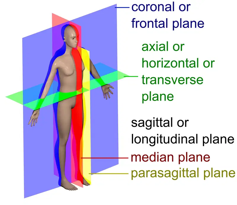
This page covers what each plane is, where the names come from, how the planes are displayed and why the patient’s left ends up on the right of your screen, how planes differ from radiographic projections, and which plane to pick for a given structure.
Axial (transverse) plane
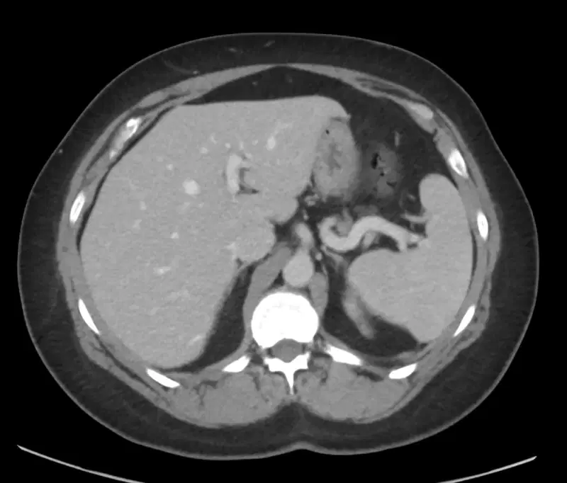
The axial plane is a horizontal plane at right angles to the long, craniocaudal axis of the body. It divides the body into a superior part and an inferior part, and an axial image is a cross section: you are looking at the cut surface of a slice, conventionally from below.
Terminologia Anatomica, the international standard for anatomical terms, lists this as the transverse plane. Axial is the radiological term, taken from the axis of the body and of the scanner gantry, and it is now so entrenched in cross-sectional imaging that the two words are used interchangeably in reports.
The distinction becomes real when the two axes stop coinciding. An axial image is perpendicular to the scanner axis, which is only a true transverse plane through a structure when that structure is aligned with the scanner. A rotated patient, a tilted gantry, or an obliquely running structure such as the pancreas, the renal artery or the femoral neck all mean that the axial slice cuts the structure obliquely, and an oblique cut through a tube always overestimates its diameter.
Head CT is the everyday example of an axial plane that is deliberately not transverse. Angling the gantry along the orbitomeatal or supraorbitomeatal baseline so that the primary beam misses the orbits cuts the dose to the lens of the eye by roughly 85 to 90 per cent, and it has traditionally also been used to reduce posterior fossa streak artefact, although no single scan plane has been shown to be clearly superior for that. A tilted head CT is therefore an angled study, and slice positions on it are not comparable with an untilted one.
Coronal (frontal) plane
The coronal plane is a vertical plane that divides the body into an anterior and a posterior part. A coronal image shows the patient as if you were facing them, which makes it the most intuitive plane for anyone who trained on radiographs, and the most efficient one for structures that run vertically: the lungs and diaphragm, the kidneys and ureters, the spine, the bowel and limb alignment.
The name comes from the Latin corona, a crown. The plane runs parallel to the coronal suture of the skull, the suture between the frontal and parietal bones that arcs across the vault from one side to the other like a tiara. Older texts sometimes claim the plane itself resembles a crown; the suture is the actual derivation.

Sagittal plane
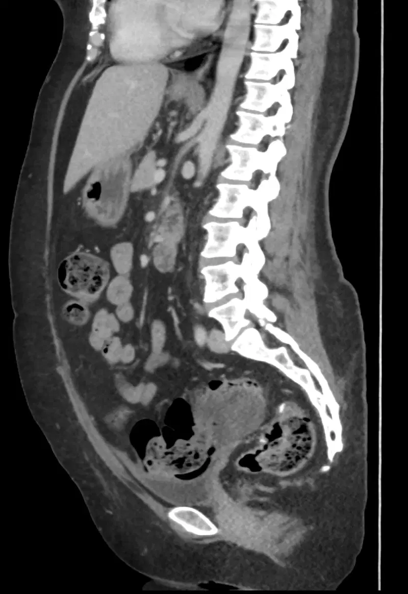
The sagittal plane is a vertical plane running front to back that divides the body into right and left parts. It is perpendicular to both the axial and the coronal planes.
Sagittal is a family of planes, not one plane, and this is where most textbook definitions go wrong. The single plane that passes exactly through the midline is the median or midsagittal plane. Any plane parallel to it but off the midline is parasagittal, also called paramedian. Saying that the sagittal plane divides the body into equal right and left halves conflates the family with its midline member.
| Term | What it means | Where you meet it |
|---|---|---|
| Sagittal plane | Any vertical front-to-back plane separating right from left | The generic term used for the whole stack of images in a sagittal series |
| Median plane (midsagittal) | The single sagittal plane through the midline | Midline sagittal T1 of the brain: corpus callosum, brainstem, pituitary fossa |
| Parasagittal (paramedian) | A sagittal plane to one side of the midline | Parasagittal meningioma; parasagittal images through a kidney or a lung hilum |
| Sagittal suture | The midline skull suture between the two parietal bones | The anatomical structure the plane is named after |
The name comes from the Latin sagitta, an arrow. The sagittal suture runs along the midline of the vault like the shaft of an arrow, meeting the lambdoid sutures posteriorly, which splay out like the flights or the arrowhead.
Etymology of the imaging planes
| Term | Root | Literal meaning | Why the plane carries the name |
|---|---|---|---|
| Axial | Latin axis | Axle, pivot | The plane is perpendicular to the long axis of the body, and to the axis of rotation of the CT gantry |
| Transverse | Latin transversus | Lying across | The plane lies across the body; this is the term used in Terminologia Anatomica |
| Coronal | Latin corona | Crown, garland | The plane runs parallel to the coronal suture, which arches across the skull vault like a crown |
| Frontal | Latin frons | Forehead, front | The synonym for coronal, reflecting the view from the front and the frontal bone |
| Sagittal | Latin sagitta | Arrow | The plane follows the sagittal suture, which runs along the vault like an arrow shaft |
| Median | Latin medianus | In the middle | The one sagittal plane that lies in the midline |
| Oblique | Latin obliquus | Slanting | Any plane angled away from the three cardinal planes, usually aligned to a structure |
Image orientation: why the patient’s left is on your right
Axial images are displayed as if you were standing at the foot of the bed, looking up towards the patient’s head. The patient’s left therefore appears on the right of the image. This is the radiological convention, and it applies to axial CT, axial MRI and body imaging generally. Coronal images follow the same rule: you are facing the patient, so their left is on your right. Sagittal images have no left-right ambiguity, but they do have an anterior-posterior one, and are conventionally displayed with the patient facing to the viewer’s left.
| Convention | Viewpoint | Patient’s left appears | Where you encounter it |
|---|---|---|---|
| Radiological | From the patient’s feet, looking cranially | On the viewer’s right | Clinical CT and MRI, PACS defaults, essentially all body imaging |
| Neurological | From above the patient’s head, looking caudally | On the viewer’s left | Some neuroimaging and research software, functional MRI and neurosurgical navigation packages |
This matters because side errors are not rare and are usually clinically important. In a review of over a million reports from a single department, side discrepancies between the body and the impression were found in 88 addended reports and a further 0.26 per cent of addended reports carried an uncorrected side error, with about 71 per cent of the errors judged clinically significant. Mirror-image reversal of coronal CT at display or printing is a described cause of wrong-side surgery in sinus and skull base practice.
- Read the L and R annotations on every study before you commit a side to the report, rather than trusting the layout on screen.
- Cross-check with an asymmetric internal landmark: the liver and gallbladder on the right, the spleen, stomach and cardiac apex on the left, the aortic arch descending on the left.
- Be suspicious when a study has been imported from outside, reformatted, screen-captured, or photographed, since each of those steps can flip an image.
- Confirm situs before applying the landmark rule; in situs inversus the landmarks reverse and the annotation is the only reliable guide.
Planes are not projections
A plane tells you where a slice was taken. A projection tells you which way the x-ray beam travelled through the patient to make a summation image. Radiographs have projections; cross-sectional images have planes. Calling a PA chest radiograph a coronal image, or a coronal reformat an AP view, muddles two different things.
| Term | What it describes | Examples | Applies to |
|---|---|---|---|
| Plane | The position and orientation of a slice through the body | Axial, coronal, sagittal, oblique, short axis | CT, MRI, ultrasound, PET, tomosynthesis |
| Projection | The direction the beam travels, from entry to exit surface | AP, PA, lateral, oblique, axial (as in the axial view of the patella or the calcaneum) | Radiography, fluoroscopy, mammography, angiography |
| View | Loose clinical shorthand, usually meaning a projection but often used for a plane | Four-chamber view, apical view, lateral view | Used across all modalities, best avoided when precision matters |
Note the trap in the word axial itself: a skyline (axial) view of the patella and an axial CT slice of the knee are named for different reasons. The first is a projection along the axis of the limb; the second is a cross section perpendicular to it.
Oblique and modality-specific planes
When a structure runs obliquely, none of the three cardinal planes cuts it cleanly, so imaging is planned along an oblique plane defined by the structure itself. These planes are prescribed at the MRI console off a scout image, or generated from a CT volume by multiplanar reformation.
| Plane | How it is defined | What it is used for |
|---|---|---|
| Cardiac short axis | Perpendicular to the long axis of the left ventricle | The basis of the American Heart Association 17-segment model, and of all perfusion and function analysis |
| Cardiac two-, three- and four-chamber (long axis) | Along the long axis of the left ventricle, rotated about it | Chamber size, valve motion, apical thrombus, regional wall motion |
| Oblique coronal shoulder | Parallel to the plane of the supraspinatus tendon and the scapular blade | Rotator cuff tendons, acromial morphology, subacromial space |
| Oblique sagittal shoulder | Perpendicular to the supraspinatus tendon | Rotator cuff muscle bulk and fatty atrophy, the rotator interval |
| Radial or oblique planes of the wrist and hip | Rotated about the axis of the joint | Triangular fibrocartilage tears, femoroacetabular impingement and the alpha angle |
| Oblique axial of the rectum or prostate | Perpendicular to the long axis of the rectum or gland | Rectal cancer staging, PI-RADS assessment of the prostate |
| Ultrasound scan planes | Defined by the probe, not by the patient | Longitudinal and transverse to the organ; transvaginal planes need their own terminology because they do not match body planes |
Ultrasound is worth singling out. The plane of a sonographic image is set by the orientation of the transducer, not by the patient’s anatomy, which is why sonographers describe planes as longitudinal and transverse to the organ. In transvaginal scanning the conventional body planes do not apply at all, and dedicated terminology has been proposed for that reason.
Acquired planes versus reformatted planes
On multidetector CT the scanner acquires a volume of near-cubic isotropic voxels in the axial plane. Coronal, sagittal and oblique images are then generated from that volume by multiplanar reformation at no extra dose and with no meaningful loss of spatial resolution. Routine coronal and sagittal reformats of the abdomen and pelvis add or change information often enough that most departments produce them by default, and coronal reformats have been judged helpful in around 60 per cent of acute abdomen studies.
MRI works the other way round. Each plane is usually a separate acquisition costing its own scan time, so planes are chosen deliberately for the clinical question, and comparative work in the thorax showed decades ago that coronal and sagittal acquisitions answer specific questions the axial stack cannot. Only 3D isotropic sequences can be reformatted freely after the fact.
| Question | First-choice plane | Why |
|---|---|---|
| Is this lesion the same on both sides | Axial | Only the axial plane places the two sides side by side at the same level |
| Where does this structure begin and end craniocaudally | Coronal | The whole length of a vertically running structure sits in one image |
| Is the bowel obstructed, and where is the transition | Coronal | Follows long loops without the reader having to track them slice to slice |
| Is there a midline lesion | Sagittal (median) | Midline structures are cut in profile rather than in cross section |
| How long is this vertebral fracture segment | Sagittal | Vertebral height loss and canal encroachment are measured in profile |
| What is the true diameter of this vessel | Oblique, perpendicular to the vessel | An oblique cut through a tube always overestimates the diameter |
Surface anatomy planes, and why the textbook levels are wrong
Transverse planes named after surface landmarks, such as the transpyloric plane, are still taught as fixed vertebral levels. Those levels come from cadaver work. When they were re-measured on CT in living supine adults, several of them moved.
| Plane | Classic teaching | CT findings in living adults |
|---|---|---|
| Sternal angle (of Louis) | T4/5 disc, with the tracheal bifurcation and aortic arch at this level | Most often T4 in women and T4/5 in men; the tracheal bifurcation, aortic arch and pulmonary trunk usually lie below the plane |
| Transpyloric plane (of Addison) | L1, at the pylorus and both renal hila | Between lower L1 and upper L2 in 75 per cent; contains the superior mesenteric artery in 56 per cent, the portal vein confluence in 53 per cent and the left renal hilum in 54 per cent, but the right renal hilum and gallbladder fundus usually lie below it |
| Subcostal plane | L3 | Most often L2 (58 per cent) |
| Supracristal plane | L4, the landmark for lumbar puncture | Most often L4 (69 per cent), the one classic level that held up well |
The practical point for reporting: quote the vertebral level you can see on the images, not the level the surface plane is supposed to correspond to.
Common mistakes
| Mistake | Correction |
|---|---|
| Calling the sagittal plane the median plane | Median (midsagittal) is the single midline sagittal plane; sagittal is the whole family |
| Calling a chest radiograph a coronal image | A radiograph is a projection, not a plane; it has no slice position and everything overlaps |
| Assuming a lesion on the right of the screen is on the patient’s right | In the radiological convention the patient’s left is on the viewer’s right; read the annotation |
| Treating an axial slice as a true transverse section of every structure | It is perpendicular to the scanner axis, not to an obliquely running organ or vessel |
| Measuring a vessel or duct on an axial image without checking its course | Measure perpendicular to the long axis of the structure, on an oblique reformat if necessary |
| Requesting a repeat scan just to obtain a coronal series on CT | Coronal and sagittal images are reformatted from the same axial volume at no extra dose |
| Quoting textbook vertebral levels for surface planes | State the level you can see; the classic levels do not hold in living supine adults |
Related reading on RadioGyan
- Radiological anatomy, a cross-sectional anatomy index organised by region.
- Normal radiology measurements, a searchable list of normal values and the plane each is measured in.
- Radiology calculators, including volume calculators that depend on measuring in three orthogonal planes.
- Radiology spotters, to practise recognising findings in every plane.
Frequently asked questions
References
- Ten Donkelaar HJ, Baud R, Kachlik D, et al. Towards a Terminologia Anatomica Humana. Anat Sci Int. 2024;99(4):387-399. PMID: 38492195
- Mirjalili SA, McFadden SL, Buckenham T, Wilson B, Stringer MD. Anatomical planes: are we teaching accurate surface anatomy? Clin Anat. 2012;25(7):819-826. PMID: 22674662
- Cerqueira MD, Weissman NJ, Dilsizian V, et al. Standardized myocardial segmentation and nomenclature for tomographic imaging of the heart. Circulation. 2002;105(4):539-542. PMID: 11815441
- Sandrasegaran K, Rydberg J, Tann M, et al. Benefits of routine use of coronal and sagittal reformations in multi-slice CT examination of the abdomen and pelvis. Clin Radiol. 2007;62(4):340-347. PMID: 17331827
- Zangos S, Steenburg SD, Phillips KD, et al. Acute abdomen: added diagnostic value of coronal reformations with 64-slice multidetector row computed tomography. Acad Radiol. 2007;14(1):19-27. PMID: 17178362
- Batra P, Brown K, Steckel RJ, et al. MR imaging of the thorax: a comparison of axial, coronal, and sagittal imaging planes. J Comput Assist Tomogr. 1988;12(1):75-81. PMID: 3335675
- Sangwaiya MJ, Saini S, Blake MA, Dreyer KJ, Kalra MK. Errare humanum est: frequency of laterality errors in radiology reports. AJR Am J Roentgenol. 2009;192(5):W239-W244. PMID: 19380530
- Schmidt D, Odland R. Mirror-image reversal of coronal computed tomography scans. Laryngoscope. 2004;114(9):1562-1565. PMID: 15475782
- Yeoman LJ, Howarth L, Britten A, Cotterill A, Adam EJ. Gantry angulation in brain CT: dosage implications, effect on posterior fossa artifacts, and current international practice. Radiology. 1992;184(1):113-116. PMID: 1609066
- Dodson MG, Deter RL. Definition of anatomical planes for use in transvaginal sonography. J Clin Ultrasound. 1990;18(4):239-242. PMID: 2160990
Image credits: anatomical planes diagram and normal CT images by David Richfield and Mikael Häggström, M.D., CC BY-SA 4.0, via Wikimedia Commons (licence).

Improve Content And Help Your Colleagues!
Found an error in the post? Please let us know using our contact page and we will update it with due credits!
If you wish to contribute radiological images for this case (or any other article on the website), you can submit them here and you will be duly credited: Submit radiology case