


Key points
- Benign cystic dilation of the rete testis, usually an incidental finding in men over 50 years; bilateral in roughly one-third of cases (reported 29-69%), often asymmetric
- Caused by obstruction at or distal to the efferent ductules at the epididymal head; commonly associated with spermatoceles and epididymal cysts
- Diagnostic confidence is highest when the process is bilateral, centered on the mediastinum testis and accompanied by epididymal cysts, with normal tumour markers
- Can present with obstructive azoospermia and infertility
- No surgery, biopsy or follow-up is required; misreading it as malignancy leads to unnecessary orchiectomy
Imaging findings
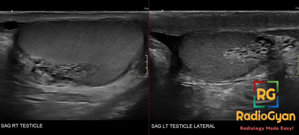
- Ultrasound: cluster of anechoic rounded and serpiginous tubular structures a few millimetres wide, centered on the mediastinum testis, without solid areas, calcification or mass effect on adjacent parenchyma
- The normal rete testis is a hypoechoic region at the testicular hilum with finger-like projections into the parenchyma; ectasia dilates these channels
- Color Doppler: no internal flow in the dilated channels
- MRI: reserved for equivocal cases; signal follows the coexisting spermatocele, hypointense to testis on T1 and proton density images and inconspicuous on T2, unlike tumours
Differential diagnosis
- Intratesticular varicocele – venous flow on Doppler that augments with Valsalva
- Cystic neoplasm, especially teratoma – solid components, calcification, mass effect, raised markers
- Cystic dysplasia of the testis – congenital, presents in children
- Epididymal cystadenoma – extratesticular, in the epididymal head; consider von Hippel-Lindau when bilateral
What to do next
- No treatment, biopsy or follow-up once the characteristic features are present
- MRI only when sonographic features are equivocal or clinical concern for malignancy persists
References
- Dogra VS, Gottlieb RH, Rubens DJ, Liao L. Benign intratesticular cystic lesions: US features. Radiographics. 2001;21 Spec No:S273-81. PMID: 11598263.
- Brown DL, Benson CB, Doherty FJ, Doubilet PM, DiSalvo DN, Van Alstyne GA, et al. Cystic testicular mass caused by dilated rete testis: sonographic findings in 31 cases. AJR Am J Roentgenol. 1992;158(6):1257-9. PMID: 1590118.
- Rouvière O, Bouvier R, Pangaud C, Jeune C, Dawahra M, Lyonnet D. Tubular ectasia of the rete testis: a potential pitfall in scrotal imaging. Eur Radiol. 1999;9(9):1862-8. PMID: 10602964.

Improve Content And Help Your Colleagues!
Found an error in the post? Please let us know using our contact page and we will update it with due credits!
If you wish to contribute radiological images for this case (or any other article on the website), you can submit them here and you will be duly credited: Submit radiology case