


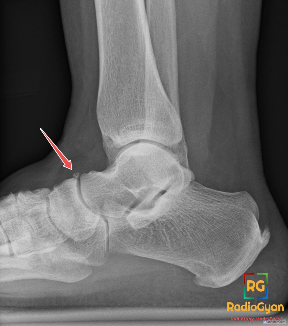
Key points
- Os supranaviculare – small accessory ossicle at the dorsal aspect of the talonavicular joint, dorsal to the navicular bone
- Also called os talonaviculare dorsalis, talonavicular ossicle or Pirie’s bone
- Rare, prevalence about 1%; usually an asymptomatic incidental finding on lateral foot and ankle radiographs
- Well-corticated margins and a matching navicular cortical edge favour ossicle over acute fracture
- Chief mimic is dorsal avulsion fracture of the talonavicular capsule – trauma history, pain and swelling, a linear irregular fragment or an associated dorsal talar head fracture favour fracture
- May fuse with the navicular forming a dorsal spur of no clinical significance; occasionally symptomatic with focal tenderness and bone marrow oedema on MRI, and reported in association with navicular stress fracture
References
- Ingalls J, Wissman R. The os supranaviculare and navicular stress fractures. Skeletal Radiol. 2011;40(7):937-41. PMID: 21484322.
- Azawi D, Mahsouli A, Vanhoenacker F. Os Supranaviculare as a Predisposing Factor to Navicular Stress Fractures. J Belg Soc Radiol. 2021;105(1):81. PMID: 34901742.

Improve Content And Help Your Colleagues!
Found an error in the post? Please let us know using our contact page and we will update it with due credits!
If you wish to contribute radiological images for this case (or any other article on the website), you can submit them here and you will be duly credited: Submit radiology case