
Quiz
Which of the following sign is NOT seen in adenomyosis?
- Rosary bead sign
- Comet Tail Sign
- Pearl Necklace Sign
- None of the above.
Answer: None of the above.
Pathophysiology
Decreased bile excretion increases intraluminal pressure in the gall bladder which leads to the proliferation of surface epithelium with outpouchings into thickened muscularis propria giving rise to intramural diverticula (also known as Rokitansky-Aschoff sinuses)
Key Imaging Features
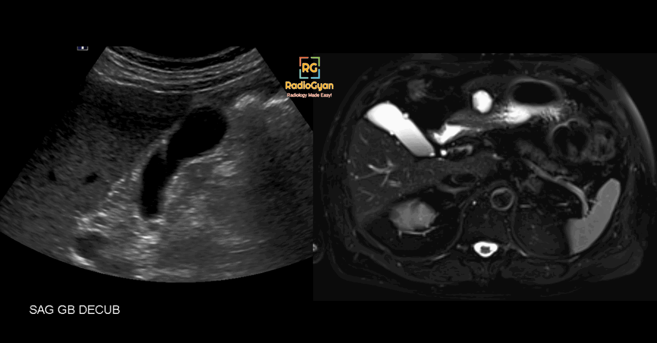
- US: Focal or segmental wall thickening with intramural echogenic foci and comet-tail reverberation artifacts as shown in the above image.
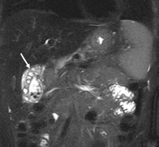
- MRI: Focal (most commonly at the fundus)or segmental wall thickening with intramural cysts (pearl necklace sign)
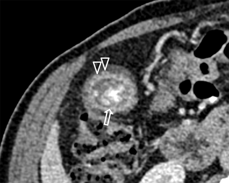
- CT: Not well seen on CT. Focal thickening at the fundus. CT rosary sign can be seen due to the intramural diverticula surrounded by the unenhanced hypertrophied muscle coat of the gallbladder.
Imaging Recommendation :
Ultrasound is diagnostic for adenomyomatosis and no further imaging is needed in most cases.
Top 3 Differential Diagnosis :
- Gallbladder Carcinoma: Infiltrative and ill-defined margin, vascular lesion, invasion into adjacent organs, and metastatic lymphadenopathy.
- Chronic Cholecystitis: Contracted gallbladder with diffuse wall thickening.
- Xanthogranulomatous Cholecystitis: Infiltrative nodular gallbladder wall thickening without comet tail artifact.
Clinical Features :
- Usually asymptomatic. Can rarely cause biliary colic.
- Not premalignant but can cause chronic GB inflammation, which is a risk factor for gall bladder malignancy.
Classification System if any :
- Focal: Most common subtype. Focal fundal thickening.
- Segmental: Circumferential thickening causing a constriction in the gallbladder. The distal portion is usually filled with calculi.
- Diffuse: Diffuse thickening with multiple intramural diverticula.
Etymology and synonyms :
- Also known as Hyperplastic Cholecystosis
- Adeno – Glands | Myoma – muscle tumors | osis – condition
Treatment :
- Cholecystectomy in symptomatic patients.
References:
Single best review article:
Other references:

Improve Content And Help Your Colleagues!
Found an error in the post? Please let us know using our contact page and we will update it with due credits!
If you wish to contribute radiological images for this case (or any other article on the website), you can submit them here and you will be duly credited: Submit radiology case