
Bosniak Calculator For Renal Cysts (CT and MRI)
What modality are you using to evalute the renal mass?
Quick References:
Other useful tools
Bosniak 2019 Renal Cyst Classification on CT and MRI
The Bosniak classification system is widely used for the characterization of renal cysts detected on imaging studies such as computed tomography (CT) and magnetic resonance imaging (MRI). This system was originally proposed by Dr. Bosniak in 1986 and was subsequently updated in 1993 and 2005. The latest update to this system, known as the Bosniak 2019 renal cyst classification, was published by Dr. Davenport in 2019.
The Bosniak classification system has become a widely accepted and essential tool for radiologists in the evaluation of renal cystic lesions. In 2019, Dr. Matthew Davenport and his team proposed an updated version of the Bosniak classification system for renal cysts on CT and MRI imaging. The latest Bosniak 2019 Renal Cyst Classification provides a more nuanced and precise approach to characterizing renal cystic lesions and helps to guide clinical management decisions, including surveillance versus intervention. The updated classification system incorporates imaging features such as the thickness of the cyst wall, septa, and calcifications to provide a more comprehensive assessment of cystic renal lesions.
This calculator is based on the original Radiology paper on the new classification by Dr. Davenport/Silverman et al. and a Radiographics paper by Dr. Schieda et al. This is a work in progress, so feedback and suggestions are welcome.
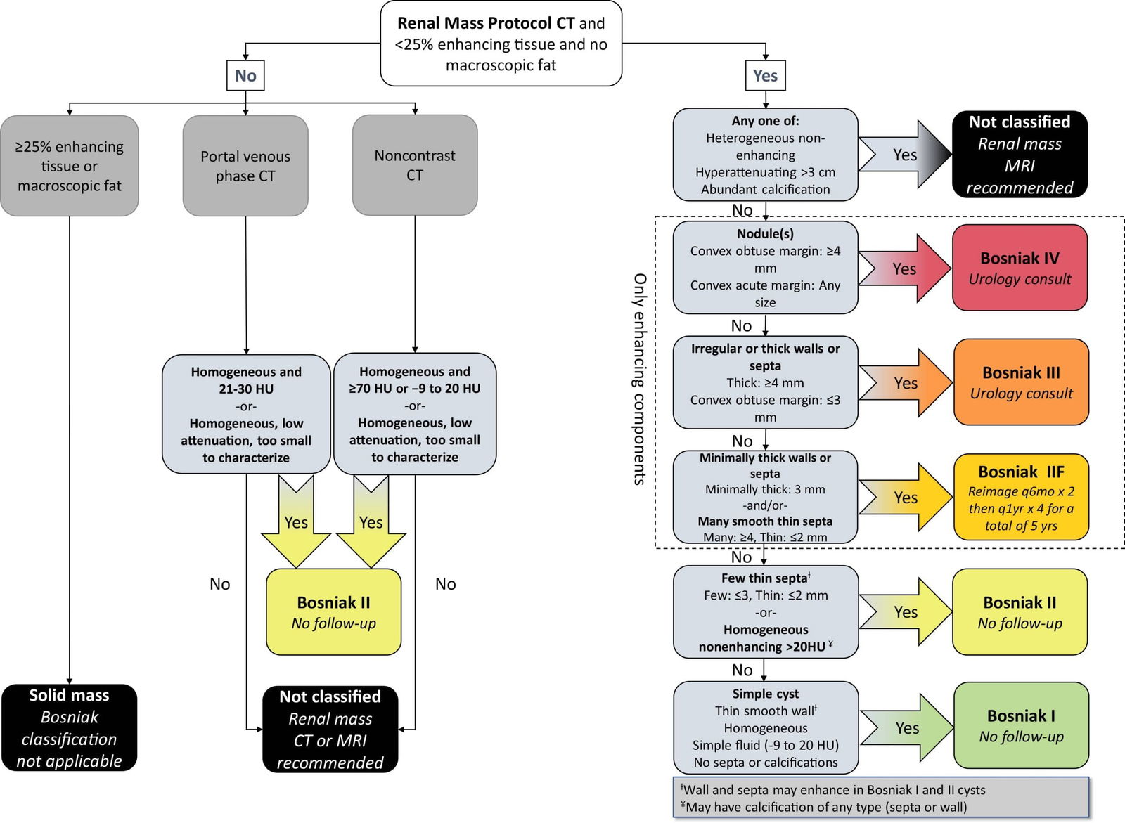
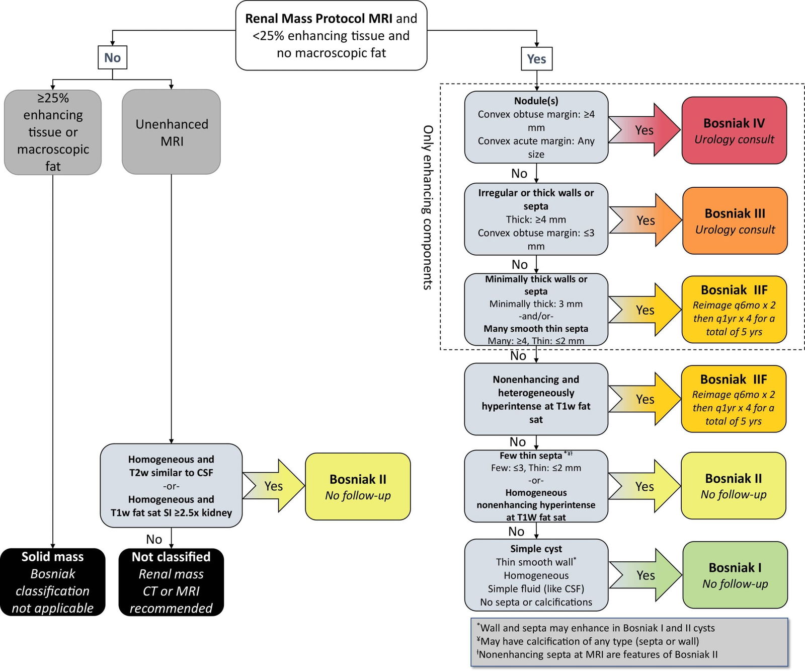
| Class | Imaging Features on CT/MRI |
|---|---|
| I | All well-defined with thin (<=2 mm) smooth walls: – Homogeneous simple fluid (-9 to 20 HU) similar. – No septa or calcifications – Wall may enhance |
| II | All well-defined with thin (<=2 mm) smooth walls: CT – Cystic masses with thin (<=2 mm) and few (1-3) septa; septa and wall may enhance; may have calcification of any type – Homogeneous hyperattenuating (>=70 HU) masses at non-contrast CT – Homogeneous nonenhancing masses > 20 HU at renal mass protocol CT, may have calcification of any type – Homogeneous masses -9 to 20 HU at noncontrast CT. – Homogeneous masses 21 to 30 HU at portal venous phase CT. – Homogeneous low attenuation masses that are too small to characterize. MRI – Homogeneous masses markedly hyperintense at T2-weighted imaging (similar to CSF) at noncontrast MRI – Homogeneous masses markedly hyperintense at T1-weighted imaging (approx 2.5 x normal parenchymal signal intensity) at noncontrast MRI |
| IIF | CT / MRI – Cystic masses with a smooth minimally thickened (3 mm) enhancing wall, or smooth minimal thickening (3 mm) of one or more enhancing septa, or many (>=4) smooth thin (<=2 mm) enhancing septa. MRI – Cystic masses that are heterogeneously hyperintense at unenhanced fat-saturated T1-weighted imaging. |
| III | One or more enhancing thick (>=4 mm width) or enhancing irregular (displaying <=3 mm obtusely marginated convex protrusions) walls or septa |
| IV | One or more enhancing nodules (>=4 mm convex protrusion with obtuse margins, or a convex protrusion of any size that has acute margins) |
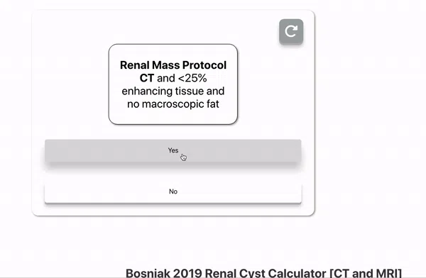
Here is a quick GIF tutorial on how to use this Bosniak Calculator
Management Recommendations for Bosniak Cysts based on CT and MRI:
| Bosniak Class | Recommendations as per 2019 Guidelines | Malignancy Rates |
|---|---|---|
| I | Benign simple renal cyst requiring no follow-up. | 0 |
| II | Likely a benign renal mass requiring no follow-up. | <1% |
| IIF | The large majority of Bosniak IIF masses are benign. When malignant, nearly all are indolent. Generally, Bosniak IIF masses are followed by imaging at 6 months and 12 months, then annually for a total of 5 years to assess for morphologic change. | 0-38% |
| III | Bosniak III masses have an intermediate probability of being malignant. If not already obtained, consider seeking a urology consultation. | ~50% |
| IV | The largest majority of Bosniak IV masses are malignant. If not already obtained, consider seeking a urology consultation. | ~90% |
Here is a quick animated tutorial for understanding the Bosniak 2019 Classification

Explore more radiology calculators on our website here:
Disclaimer: The author makes no claims of the accuracy of the information contained herein; this information is for educational purposes only and is not a substitute for clinical judgment.
References:
- Bosniak Classification of Cystic Renal Masses, Version 2019: An Update Proposal and Needs Assessmen. Stuart G. Silverman, Ivan Pedrosa, James H. Ellis, Nicole M. Hindman, Nicola Schieda, Andrew D. Smith, Erick M. Remer, Atul B. Shinagare, Nicole E. Curci, Steven S. Raman, Shane A. Wells, Samuel D. Kaffenberger, Zhen J. Wang, Hersh Chandarana, and Matthew S. DavenportRadiology 2019 292:2, 475-488
- Bosniak Classification of Cystic Renal Masses, Version 2019: A Pictorial Guide to Clinical Use. Nicola Schieda, Matthew S. Davenport, Satheesh Krishna, Elizabeth A. Edney, Ivan Pedrosa, Nicole Hindman, Ronaldo H. Baroni, Nicole E. Curci, Atul Shinagare, and Stuart G. SilvermanRadioGraphics 2021 41:3, 814-828

Improve Content And Help Your Colleagues!
Found an error in the post? Please let us know using our contact page and we will update it with due credits!
If you wish to contribute radiological images for this case (or any other article on the website), you can submit them here and you will be duly credited: Submit radiology case
About the Author
Dr. Amar Udare, MD, DNB
Dr. Udare holds an MBBS and MD degree, and his expertise lies in the field of radiology. He has authored multiple peer-reviewed publications, contributing significantly to the medical field. His works can be accessed on PubMed and Google Scholar.
In addition to his academic and professional achievements, Dr. Udare is an avid reader and enjoys exploring the latest advancements in medical technology. His commitment to making complex medical knowledge accessible to patients and the general public aligns with our mission at RadioGyan.com.
For any further questions or clarifications, feel free to reach out to Dr. Udare via the contact form.