


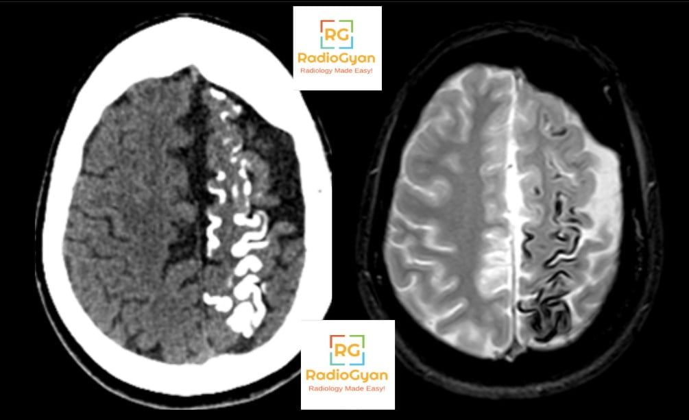
Sturge-Weber syndrome is a sporadic capillary-venous malformation syndrome of the skin, brain and eye, caused by a somatic activating mutation in GNAQ (most often p.R183Q) that arises after fertilisation, so the child is a genetic mosaic and nothing is inherited [7]. Current consensus defines it as at least two of a facial capillary malformation, a cerebral vascular malformation and ocular involvement [2] — which means a child can have Sturge-Weber syndrome with no port-wine birthmark at all.
Two things about the imaging have changed enough since the classical description to matter in daily reporting. First, the findings most textbooks list — pial enhancement, tram-track calcification, atrophy — are late, and a normal-looking scan in an infant does not exclude the diagnosis. Second, the lesion itself is not an angioma. This page covers the mnemonic, the early and late imaging findings separately, who should be scanned and when, and where the classical teaching is now wrong.
Key points
- Sturge-Weber syndrome needs two of three: facial capillary malformation, cerebral vascular malformation, ocular involvement [2].
- The brain lesion is a subarachnoid varicose network of dilated veins compensating for absent cortical veins. “Leptomeningeal angioma” and “pial angiomatosis” are misnomers that should be retired [1].
- Pial-arachnoid enhancement is often not visible before 1 year of age; a negative early MRI does not exclude the diagnosis [1,3].
- Early, non-contrast markers carry the diagnosis in infancy: choroid plexus thickness 5.6 mm or more on the affected side (about 92 percent sensitive, 100 percent specific), enlarged deep medullary veins on SWI, and asymmetric accelerated myelination [1,5].
- Tram-track calcification is a late, insensitive finding. SWI beats CT for detecting it [1].
- Risk of brain involvement is predicted by a birthmark involving any part of the forehead, following embryonic facial vasculature rather than the trigeminal divisions [4].
- Arterial spin labelling shows hyperperfusion early and hypoperfusion late; the direction of the perfusion abnormality dates the disease [1,9].
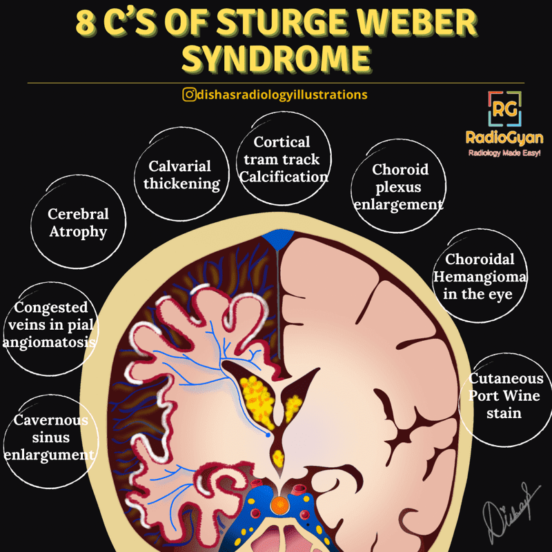
The 8 C’s mnemonic for Sturge-Weber syndrome
| The C | What it means | How it looks |
|---|---|---|
| Cutaneous port-wine birthmark | Facial capillary malformation, the cutaneous hallmark | Clinical. Risk of brain involvement rises with forehead and upper-eyelid involvement [4] |
| Congested subarachnoid veins | The dilated venous network in the subarachnoid space, formerly miscalled pial angiomatosis | Pial-arachnoid enhancement on post-contrast T1 and post-contrast FLAIR; serpiginous flow voids on T2 [1] |
| Choroid plexus enlargement | Congested plexus from diverted deep venous drainage | Ipsilateral, measurable on non-contrast T2 or T1. Cut-off 5.6 mm on the affected side [5] |
| Cortical tram-track calcification | Gyriform cortical and subcortical calcification from chronic venous ischaemia | Parallel gyriform lines on CT and radiographs; blooming on SWI or gradient echo [1] |
| Cerebral atrophy | Progressive volume loss in the affected territory | Hemispheric or lobar, usually parieto-occipital, with widened sulci |
| Calvarial thickening | Compensatory Dyke-Davidoff-Masson type change to the shrinking hemisphere | Ipsilateral diploic thickening, elevated petrous ridge, hyperpneumatised sinuses and mastoid |
| Choroidal haemangioma | Diffuse choroidal vascular malformation in the eye | Diffuse choroidal thickening on ocular ultrasound, MRI or OCT; occurs in roughly 40 to 50 percent |
| Chronically raised intraocular pressure | Glaucoma, in 30 to 70 percent, and buphthalmos when it starts in infancy | Clinical and tonometric. Needs lifelong ophthalmology follow-up [2] |
One correction to the classical list. Older versions of this mnemonic include “cavernous sinus enlargement”. That is not a recognised Sturge-Weber feature. The venous structures that enlarge are the choroid plexus, the deep medullary veins and the subependymal veins, all of them part of the centripetal drainage that takes over when the cortical veins fail [1]. The eighth C is better spent on the choroid plexus.
Roach classification and where it falls short
| Roach type | Facial capillary malformation | Brain involvement | Glaucoma |
|---|---|---|---|
| Type I (classic) | Present | Present | May be present |
| Type II | Present | Absent | May be present |
| Type III | Absent | Present (isolated) | Usually absent |
The Roach scale is still the vocabulary of most reports, but it is now regarded as oversimplified, particularly for Type II, because it treats brain involvement as a binary and ignores the fact that the risk is stratified by the size and site of the birthmark [1]. The 2025 ISSVA classification instead places the condition among the syndromic slow-flow port-wine capillary malformations. In practice, use Roach for communication and describe the phenotype in full.
Early versus late MRI findings
This is the section that changes reporting. The disease is a progressive venous congestion, so imaging findings move through a sequence, and the ones every textbook illustrates sit at the end of it. Seizures usually begin before the age of 2, which means the diagnostically useful window is the one in which the classical findings are still absent [1].
| Stage | Finding | Sequence | Why it happens |
|---|---|---|---|
| Early (neonate to about 1 year) | Enlarged deep medullary veins; subarachnoid varicose network | SWI, T2 | Cortical veins absent or dysplastic; flow redirected into the deep system. Reported as early as 5 to 11 days of life [1] |
| Early | Choroid plexus enlargement, 5.6 mm or more on the affected side | Non-contrast T1 or T2 | Subependymal veins drain the plexus; once overwhelmed the plexus congests. About 92 percent sensitive, 100 percent specific [5] |
| Early | Accelerated myelination: asymmetric low T2 signal, high T1 signal in subcortical white matter | T1, T2 | Transient hyperperfusion and hypermetabolism. Usually identifiable before 6 months [1] |
| Early | Hyperperfusion of the affected cortex | Arterial spin labelling | Compensatory phase, before tissue loss. Seen in younger, asymptomatic patients with little atrophy [1,9] |
| Established | Pial-arachnoid enhancement following sulci and cisterns | Post-contrast T1, post-contrast FLAIR | The subarachnoid varicose network. First reported as early as 35 days but frequently not visible before 1 year [1] |
| Late | Gyriform cortical and subcortical calcification (tram-track) | SWI, gradient echo, CT | Chronic ischaemic cortical injury. Accumulates with age [1] |
| Late | Cortical laminar necrosis, gliosis, lobar or hemispheric atrophy | T1, FLAIR, DWI | Irreversible injury from sustained venous congestion |
| Late | Hypoperfusion of the affected cortex | Arterial spin labelling | Burn-out stage; associated with extensive calcification and severe epilepsy [1] |
| Late | Ipsilateral calvarial thickening, sinus and mastoid hyperpneumatisation, elevated petrous ridge | CT, T1 | Compensatory change to the atrophic hemisphere (Dyke-Davidoff-Masson pattern) |
Suggested MRI protocol
- 3.0 T where available, with sedation planned rather than improvised. In infants a feed-and-wrap, non-contrast study is a reasonable first pass [2].
- Volumetric T1 pre- and post-contrast for pial-arachnoid enhancement and for the choroid plexus.
- SWI or T2* — the single highest-yield sequence. It shows the deep medullary veins, the subarachnoid varicose network and calcification, all without contrast [1].
- Arterial spin labelling, acquired before gadolinium, with a tailored post-label delay. Interpret with caution close to a seizure, since ictal and post-ictal perfusion changes mimic disease [1].
- Post-contrast FLAIR is more sensitive than post-contrast T1 for subtle leptomeningeal enhancement.
- T2 and DWI for myelination pattern, gliosis and acute stroke-like injury.
- Venous MR angiography and high-resolution orbital sequences are included in the 2025 consensus protocol [2].
Why leptomeningeal angioma is the wrong name
Radio-pathological correlation finds no angioma. What is present is a network of dilated, varicose veins in the subarachnoid space, compensating for absent or dysplastic superficial cortical drainage. The 2025 Pediatric Radiology review states the term is outdated and should be avoided, preferring subarachnoid varicose network or leptomeningeal capillary-venous malformation [1].
This is not pedantry, because the name drives the mental model. “Angioma” implies a proliferative mass lesion and invites questions about haemorrhage and resection. “Venous congestion” explains, in one mechanism, the whole imaging sequence: diverted deep venous drainage gives choroid plexus enlargement and enlarged medullary veins; early congestion gives hyperperfusion and accelerated myelination; sustained congestion gives ischaemia, calcification, laminar necrosis, atrophy and finally hypoperfusion. It also explains why these are low-flow malformations that do not need catheter angiography unless surgery is planned.
Which birthmarks to work up, and when to scan
The old teaching that the birthmark follows the V1 trigeminal division is wrong. In 192 children with a facial port-wine stain, the strongest predictor of seizures, abnormal neurodevelopment, glaucoma and abnormal MRI was involvement of any part of the forehead, its lower border a line from the outer canthus of the eye to the top of the ear, including the upper eyelid. That area crosses all three trigeminal divisions but corresponds closely to the embryonic vasculature of the face. Bilateral distribution was not independently significant [4].
| Question | US consensus, 2021 [3] | Italian consensus, 2025 [2] |
|---|---|---|
| Which birthmark triggers workup | High-risk facial port-wine birthmark: refer to paediatric neurology and paediatric ophthalmology for baseline assessment and periodic follow-up | Frontal region bordered inferiorly by outer canthus to upper ear, including upper eyelid, plus midline frontonasal. About 81 percent of children with that distribution had the syndrome in the cited series |
| Screening MRI in an asymptomatic infant | Not routinely recommended. May be performed in selected cases, for example where presymptomatic treatment is being considered | Recommended, preferably after 12 months, with gadolinium. Non-contrast feed-and-wrap within 4 weeks if suspicion is high |
| Repeat imaging | Routine follow-up neuroimaging not recommended if symptoms are stable | If negative after 12 months and the child stays asymptomatic, wait to around age 6 to avoid repeat sedation |
| EEG | Not specified | Baseline sleep EEG in the first 3 months for high-risk birthmarks; 6-monthly for the first 2 years once confirmed |
| Ophthalmology | Baseline evaluation and periodic follow-up; intervals not defined | Complete examination at diagnosis, then annual review from birth if uncomplicated; tonometry under anaesthesia until the child cooperates; lifelong review in adults |
| Presymptomatic drugs | Retrospective data suggest benefit from antiseizure medication plus aspirin; no completed prospective trial | Antiseizure medication may be offered where brain involvement is extensive and early-onset epilepsy risk is high, with the empirical basis explained to the family |
The disagreement is real and worth stating in a report or a multidisciplinary discussion rather than papering over. It comes down to the same fact read two ways: a negative MRI before the first birthday does not exclude the diagnosis. Negative neuroimaging in infants with a high-risk birthmark who later proved to have the syndrome was reported in 3 to 23 percent of retrospective cases [3], and the leptomeningeal malformation was undetectable on the initial scan in 21 percent of children imaged at 3 months or younger [5]. The US group concludes that an unreliable test should not be done routinely; the Italian group concludes it should be delayed until it becomes reliable. What rescues the early scan is the indirect signs: a direct or at least one indirect sign on a first MRI before 3 months gave 100 percent sensitivity and 94 percent specificity for the diagnosis [6].
Ocular involvement
- Glaucoma in 30 to 70 percent, with a bimodal onset — infancy, from a maldeveloped anterior chamber angle, and later childhood or adulthood, from raised episcleral venous pressure. Infantile onset produces buphthalmos, an enlarged globe visible on any brain MRI.
- Diffuse choroidal haemangioma in roughly 40 to 50 percent, ipsilateral to the birthmark. On MRI it is diffuse choroidal thickening that enhances; it is the cause of the “tomato ketchup fundus” and can cause exudative retinal detachment and vision loss.
- Episcleral and conjunctival vascular lesions and dilated tortuous retinal vessels.
- Look for these on every brain MRI in a child with a port-wine birthmark. Globe asymmetry and choroidal thickening are easy to see and easy to skip past.
Differential diagnosis
| Entity | What overlaps | What separates it |
|---|---|---|
| PHACE syndrome | Segmental facial vascular lesion with intracranial abnormality | The skin lesion is an infantile haemangioma, not a capillary malformation, and the intracranial findings are posterior fossa malformation, arterial anomalies, coarctation and eye anomalies |
| Meningioangiomatosis | Cortical and leptomeningeal lesion with gyriform calcification and seizures | Focal, often frontal or temporal, associated with neurofibromatosis type 2; no birthmark, no hemispheric atrophy pattern |
| Klippel-Trenaunay syndrome | Capillary malformation, sometimes PIK3CA-related | Limb capillary-lymphatic-venous malformation with overgrowth; no leptomeningeal disease |
| Coeliac disease with epilepsy and occipital calcification | Bilateral occipital gyriform calcification with seizures | No enhancement, no birthmark, no atrophy of the classical Sturge-Weber pattern; gluten enteropathy |
| Cortical laminar necrosis from hypoxic-ischaemic injury | Gyriform T1 hyperintensity and later calcification | Follows a vascular or watershed distribution with a clear insult; no leptomeningeal enhancement |
| Congenital infection (CMV, toxoplasmosis) | Intracranial calcification with atrophy and seizures | Periventricular or basal ganglia calcification, migrational anomaly, microcephaly; not gyriform and cortical |
| Dyke-Davidoff-Masson syndrome | Hemiatrophy with ipsilateral calvarial thickening and sinus hyperpneumatisation | The compensatory skull change is shared. Sturge-Weber adds enhancement, gyriform calcification and the birthmark; Dyke-Davidoff-Masson is the end-state of any early hemispheric insult |
Management in brief
| Problem | Treatment | Evidence |
|---|---|---|
| Seizures | Standard antiseizure medication; levetiracetam and oxcarbazepine are the most used. Drug-resistant epilepsy: focal resection, hemispherotomy or vagus nerve stimulation | Cross-sectional multicentre survey data; surgery reserved for medically refractory cases [8] |
| Stroke-like episodes | Low-dose aspirin, 3 to 5 mg/kg/day up to 100 mg | Offered by many but not all centres; retrospective data suggest fewer stroke-like episodes and seizures. Offered early once neurological symptoms appear in extensive disease [2,8] |
| Presymptomatic prevention | Antiseizure medication plus aspirin before the first seizure, in selected infants with extensive brain involvement | May delay seizure onset; hypothesis-generating retrospective data, no completed prospective trial [3,10] |
| Cognition | Sirolimus | Prospective open-label trial showing a signal for improved processing speed [11] |
| Refractory seizures | Cannabidiol | Prospective trial support [8] |
| Port-wine birthmark | Pulsed dye laser, starting in the first year of life, sessions 4 to 6 weeks apart | Earlier treatment gives better outcomes; sedation or general anaesthesia preferred under 1 year to avoid pain memory [2] |
| Glaucoma | Medical intraocular pressure control, then angle or filtration surgery; lifelong monitoring | Tonometry under anaesthesia until the child cooperates; visual field testing in adults [2] |
What to put in the report
- Which lobes and which hemisphere, and whether involvement is unilateral or bilateral. Bilateral disease predicts earlier seizures and worse neurodevelopment.
- Pial-arachnoid enhancement: present or absent, and its extent along sulci. If the child is under 1 year and the study is negative, say explicitly that absent enhancement does not exclude the diagnosis at this age [1,3].
- Choroid plexus thickness on each side, in millimetres. This is a measurement, not an impression, and 5.6 mm or more on the affected side is the published threshold [5].
- Deep medullary and subependymal vein prominence on SWI, and any subarachnoid varicose network.
- White matter signal: asymmetric accelerated myelination early, gliosis and volume loss later.
- Calcification and its distribution, ideally described on SWI rather than requesting a CT.
- Perfusion: hyper- or hypoperfused, and the caveat that a recent seizure confounds it.
- Atrophy and the compensatory skull changes, with a comparison to any prior study, since progression drives surgical decisions.
- The orbits: globe size asymmetry and diffuse choroidal thickening.
Related reading on RadioGyan
- Tram-track sign — the sign itself, and the other conditions that produce it.
- Von Hippel-Lindau syndrome — the other classic phakomatosis, with its surveillance schedule.
- PHACE syndrome — the segmental facial vascular lesion that is not a capillary malformation.
- Neurofibromatosis — imaging findings across types 1 and 2.
- Illustrated radiology mnemonics — the full collection.
Frequently asked questions
References
- Cerron-Vela CR, Manteghinejad A, Andronikou S. Beyond the leptomeningeal angioma: a comprehensive review of MR imaging features of Sturge-Weber syndrome, from early vascular responses to tissue necrosis. Pediatr Radiol. 2025;55(13):2704-2715. PMID 41137931
- El Hachem M, Diociaiuti A, Latorre S, Ficcadenti A, Kiener A, Marinelli F, et al. Multidisciplinary, multicenter consensus for the care of patients affected with Sturge-Weber syndrome. Orphanet J Rare Dis. 2025;20(1):28. PMID 39819452
- Sabeti S, Ball KL, Bhattacharya SK, Bitrian E, Blieden LS, Brandt JD, et al. Consensus statement for the management and treatment of Sturge-Weber syndrome: neurology, neuroimaging, and ophthalmology recommendations. Pediatr Neurol. 2021;121:59-66. PMID 34153815
- Waelchli R, Aylett SE, Robinson K, Chong WK, Martinez AE, Kinsler VA. New vascular classification of port-wine stains: improving prediction of Sturge-Weber risk. Br J Dermatol. 2014;171(4):861-867. PMID 24976116
- Catsman-Berrevoets CE, Koudijs SM, Buijze MSJ, de Laat PCJ, Pasmans SGMA, Dremmen MHG. Early MRI diagnosis of Sturge Weber syndrome type 1 in infants. Eur J Paediatr Neurol. 2022;38:66-72. PMID 35461064
- Bar C, Pedespan JM, Boccara O, Garcelon N, Levy R, Grevent D, et al. Early magnetic resonance imaging to detect presymptomatic leptomeningeal angioma in children with suspected Sturge-Weber syndrome. Dev Med Child Neurol. 2020;62(2):227-233. PMID 31050360
- Shirley MD, Tang H, Gallione CJ, Baugher JD, Frelin LP, Cohen B, et al. Sturge-Weber syndrome and port-wine stains caused by somatic mutation in GNAQ. N Engl J Med. 2013;368(21):1971-1979. PMID 23656586
- Yeom S, Comi AM. Updates on Sturge-Weber syndrome. Stroke. 2022;53(12):3769-3779. PMID 36263782
- Pouliquen G, Fillon L, Dangouloff-Ros V, Kuchenbuch M, Bar C, Chemaly N, et al. Arterial spin-labeling perfusion imaging in the early stage of Sturge-Weber syndrome. AJNR Am J Neuroradiol. 2022;43(10):1516-1522. PMID 36137664
- Day AM, Hammill AM, Juhasz C, Pinto AL, Roach ES, McCulloch CE, et al. Hypothesis: presymptomatic treatment of Sturge-Weber syndrome with aspirin and antiepileptic drugs may delay seizure onset. Pediatr Neurol. 2019;90:8-12. PMID 30482419
- Sebold AJ, Day AM, Ewen J, Adamek J, Byars A, Cohen B, et al. Sirolimus treatment in Sturge-Weber syndrome. Pediatr Neurol. 2021;115:29-40. PMID 33316689
- Ramirez EL, Julich K. Sturge-Weber syndrome: an overview of history, genetics, clinical manifestations, and management. Semin Pediatr Neurol. 2024;51:101151. PMID 39389653
Causes Of Cerebral Rim Enhancing Lesions – Visual Mnemonics

Hepatic Cystic Lesions- Visual Mnemonics
Causes of Apical Lung Fibrosis – Visual Mnemonics

Improve Content And Help Your Colleagues!
Found an error in the post? Please let us know using our contact page and we will update it with due credits!
If you wish to contribute radiological images for this case (or any other article on the website), you can submit them here and you will be duly credited: Submit radiology case
Thank you very much doctors your educational site is magnificent.
Glad you liked it!