
Introduction
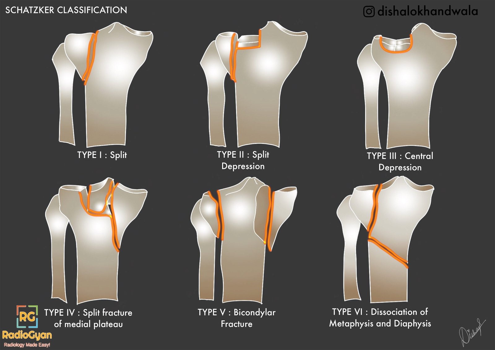
The Schatzker classification system for tibial plateau fractures helps orthopedic surgeons to assess the initial injury, plan management, and predict prognosis. It was initially described on radiographs but CT and MRI are more accurate. There are six fracture types in increasing order of severity and worse prognosis.
Schatzker Classification of tibial plateau fractures
Type I Lateral plateau fracture without depression or depression <4mm
Type II Lateral plateau fracture with depression
Type III Pure depression fracture – IIIA Lateral; IIIB – Central depression
Type IV Medial plateau fracture
Type V Bicondylar fracture
Type VI Plateau fracture with diaphyseal discontinuity.
Types I-III are low-energy injuries while IV-VI are high-energy injuries
Illustration
Characteristics of individual Schatzker fracture types
| Type | Characteristics |
|---|---|
| I | – Wedge-shaped pure cleavage fracture of the lateral tibial plateau – 6% of all tibial plateau fractures – More frequent in young patients with normal bone mineralization – May be subtle on radiographs and confused with type II fracture. – May be associated with type distraction type MCL / ACL injury. – Treatment: Open reduction and internal fixation with or without arthroscopy. |
| II | – Combined cleavage and compression fracture of the lateral tibial plateau, a type I fracture with a depressed component. – Depression may be difficult to appreciate on plain radiographs. – Depression is measured as the vertical distance between the lowest point on the intact medial plateau and the lowest depressed lateral plateau fracture fragment. – 25% of all tibial plateau fractures. – Common in the 4th decade. – Associated distraction injuries to the MCL or medial meniscus – Treatment: Open reduction |
| III | – Pure compression fracture of the lateral (IIIA) or central(IIIB) tibial plateau. – Articular surface depressed into metaphysis by axial forces. – Account for 36% of cases. – Common in the 4th to 5th decade. – Treatment: IIIA – May be non-operative. IIIB – Depressed portion of the plateau is typically elevated by means of a submetaphyseal cortical window |
| IV | – Medial tibial plateau fracture with a split or depressed component. – 10% of all plateau fractures. – Poor prognosis. – Associated with injury to the peroneal nerve or popliteal vessels and lateral collateral ligament / posterolateral ligament complex. – Treatment: Open reduction and internal fixation. |
| V | – Bicondylar wedge fracture with an inverted Y appearance. – Only 3 % of all tibial fractures. – Associated with high-energy trauma. ACL and meniscal involvement common. – Collateral ligament anchors are disrupted, the ligaments themselves may be intact. – Initial management involves splinting or temporary external fixation to allow for soft-tissue inflammation and edema to subside before surgical management. |
| VI | – Transverse subcondylar fracture with dissociation of the metaphysis from the diaphysis. – 20% of all tibial plateau fractures. – High energy trauma and associated with an extensive soft-tissue injury and increased risk of compartment syndrome. |
Case Example

The above fracture is a type II tibial plateau fracture. Click here for more images.
References and further reading
Illustration contributed by #TeamGyan member Dr. Disha Lokhandwala.

Improve Content And Help Your Colleagues!
Found an error in the post? Please let us know using our contact page and we will update it with due credits!
If you wish to contribute radiological images for this case (or any other article on the website), you can submit them here and you will be duly credited: Submit radiology case