


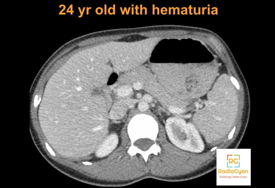
Nutcracker syndrome is symptomatic compression of the left renal vein, usually between the aorta and the superior mesenteric artery, causing left flank pain and haematuria. On imaging, the most reliable findings are a beak sign with a hilar to aortomesenteric diameter ratio of 4.9 or more on CT, and a peak velocity ratio above about 5 on Doppler ultrasound. The trap is that compression without symptoms, the nutcracker phenomenon, is common. Nearly a third of healthy kidney donors have an aortomesenteric angle under 41 degrees [1], so a CT finding alone should be reported as nutcracker anatomy, not as the syndrome.
This article is a working reference for reporting a suspected nutcracker: the Doppler, CT, MRI and venographic criteria with their real sensitivities and specificities, how often healthy people meet each one, anterior versus posterior nutcracker, the differential diagnosis, and what the 2025 international Delphi consensus did and did not agree [2]. It ends with a reporting checklist and the post-treatment findings you will be asked to assess.
- Key facts for practice and radiology board exams
- Nutcracker phenomenon versus nutcracker syndrome
- Anatomy: anterior and posterior nutcracker
- Clinical presentation
- Doppler ultrasound criteria
- CT criteria
- MRI
- Venography, pressure gradient and IVUS
- What the 2025 Delphi consensus agreed
- Where nutcracker fits in the SVP classification
- Differential diagnosis
- Treatment and post-treatment imaging
- How to report a suspected nutcracker
- Frequently asked questions
- References
Key facts for practice and radiology board exams
- Phenomenon versus syndrome. Compression without symptoms is nutcracker phenomenon (nutcracker anatomy). Syndrome needs compatible symptoms, and the 2025 Delphi panel agreed they should last more than 6 months [2].
- Commonest features: haematuria 69.5 percent, left flank or abdominal pain 48.4 percent, pelvic pain 23.1 percent, varicocele 15.8 percent in a 384-patient systematic review [3]. In children, haematuria and proteinuria dominate and flank pain is present in only 19.1 percent [4].
- CT: beak sign 91.7 percent sensitive and 88.9 percent specific against venographic gradients; the beak sign plus a diameter ratio of 4.9 or more is the most accurate combination [5].
- Aortomesenteric angle under 41 degrees: pooled sensitivity 94 percent, specificity 85 percent [6], but present in 30.5 percent of healthy donors [1]. Sensitive, not specific in practice.
- Doppler: measure diameter and peak velocity at the hilum and at the aortomesenteric segment. Peak velocity ratio averaged 7.9 in patients versus 2.8 in controls [7]; ratios above 4 (diameter) and 5 (velocity) are the usual thresholds [8].
- Posterior nutcracker = retroaortic left renal vein compressed between aorta and vertebra. The variant is present in 0.77 percent of CT scans and symptomatic in only 6.6 percent of those [9].
- No gold standard. The Delphi panel agreed there is none, did not agree that phlebography is the gold standard, and could not agree on any imaging cut-off [2].
- Normal renocaval gradient is 0 to 1 mm Hg; 3 mm Hg or more indicates non-compensated nutcracker syndrome [5]. A normal gradient does not exclude the syndrome, because collaterals decompress the vein [2].
- Treatment: conservative first. The first-choice operation is left renal vein transposition, and endovascular stenting is not recommended as primary treatment because of stent migration [2].
Nutcracker phenomenon versus nutcracker syndrome
Every criterion in this article describes the anatomy, not the illness. The 2025 Delphi panel, 20 venous experts from the Americas, Europe and Asia-Pacific, agreed that nutcracker syndrome is a combination of clinical signs and symptoms caused by stenosis of the left renal vein (100 percent agreement), that its characteristic features are haematuria and flank pain (100 percent), and that the discovery of asymptomatic compression is the nutcracker phenomenon (85 percent) [2]. They ranked flank pain as the most relevant symptom, followed by haematuria, pelvic pain and proteinuria.
The reason the distinction matters to a radiologist is prevalence. In 99 CT angiograms of potential renal donors, 23 had 50 to 70 percent left renal vein stenosis and 4 had more than 70 percent, and neither the compression nor dilated gonadal and lumbar veins were associated with haematuria or pain [10]. In 324 healthy donors, the beak sign was present in 15.3 percent and an aortomesenteric angle under 41 degrees in 30.5 percent, both more often in women and in younger people [1]. If you write “nutcracker syndrome” in the conclusion of a CT done for something else, you have almost certainly overcalled it.
| Nutcracker phenomenon (anatomy) | Nutcracker syndrome | |
|---|---|---|
| Definition | Imaging compression of the left renal vein, no attributable symptoms | Compression plus compatible symptoms, lasting more than 6 months [2] |
| How often | Common: 27 of 99 donors had 50 percent or more stenosis [10] | Rare; no reliable population prevalence |
| Typical setting | Incidental on CT for another indication; commoner in women and younger people [1] | Flank pain, haematuria, orthostatic proteinuria, left varicocele, pelvic venous symptoms |
| What to report | Describe the compression and measurements; use the words nutcracker anatomy | Describe compression, measurements, collaterals and gonadal reflux; suggest duplex correlation or venography |
| Treatment | None | Conservative first; intervention for persistent or severe symptoms |
Anatomy: anterior and posterior nutcracker
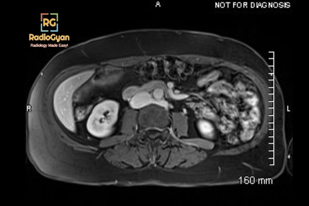
The left renal vein normally crosses anterior to the aorta, just below the origin of the superior mesenteric artery, to reach the inferior vena cava. In anterior nutcracker it is squeezed in the fork between the two arteries; in posterior nutcracker a retroaortic left renal vein is compressed between the aorta and the vertebral body. Renal venous hypertension drains through collaterals: the left gonadal vein (pelvic varices, left varicocele), the ascending lumbar and hemiazygos system, ureteric and capsular veins, and renal hilar varices.
| Anterior nutcracker | Posterior nutcracker | |
|---|---|---|
| Compressing structures | Aorta and superior mesenteric artery | Aorta and vertebral body |
| Venous anatomy | Normal pre-aortic left renal vein | Retroaortic left renal vein (or retroaortic limb of a circumaortic vein) |
| How common | The usual form | Retroaortic vein in 0.77 percent of 7,929 CTs; 6.6 percent of those symptomatic [9] |
| Key image | Sagittal reformat showing an acute aortomesenteric angle; axial beak sign | Axial image showing the vein flattened behind the aorta against the spine |
| Presentation | Flank pain, haematuria, proteinuria, varicocele, pelvic pain | Haematuria in 25 and flank pain in 13 of 27 published cases [11] |
| Usual management | Conservative first; transposition or stenting if refractory | Conservative in most cases [11] |
Clinical presentation
| Feature | Adults and mixed series | Children |
|---|---|---|
| Haematuria | 69.5 percent [3] | 55.5 percent [4] |
| Left flank or abdominal pain | 48.4 percent [3] | 19.1 percent [4] |
| Proteinuria | Less common | 49.9 percent [4]; renal nutcracker found in 68 percent of 229 children with orthostatic proteinuria [12] |
| Pelvic pain | 23.1 percent [3] | Uncommon |
| Varicocele | 15.8 percent [3] | Nutcracker phenomenon in 56 percent of adolescents with a varicocele [13] |
Orthostatic proteinuria in children is the setting where the natural history is most benign: in a group reassessed 6 years after diagnosis, both the proteinuria and the compression had disappeared in 9 of 13 [12]. The panel could not agree on the mechanism of haematuria or proteinuria [2].
Doppler ultrasound criteria
Duplex ultrasound is the first test. The 2025 Delphi panel agreed that it should be part of the workup (90 percent), that a report should at least give the peak systolic velocity at the stenosis and in the distal renal vein and the vein diameter at and distal to the stenosis (90 percent), and that because of its variability it should not be the only modality used to make the diagnosis (100 percent) [2].
Technique. Measure at two points: the renal hilum, and the aortomesenteric segment where the vein crosses between the aorta and the superior mesenteric artery. Correct the Doppler angle and keep the sample volume small at the narrowing; these are the main technical pitfalls [14]. Scan supine and then standing or sitting: compression increases in the upright position, and the standing velocity ratio was the most accurate single parameter in a 52-patient series [15].
| Parameter | Threshold | Performance | Source |
|---|---|---|---|
| Peak velocity ratio (aortomesenteric to hilar) | Above about 5 | Mean 7.9 in 16 patients versus 2.8 in 18 controls | [7] |
| Anteroposterior diameter ratio (hilar to aortomesenteric) | Above about 4 to 5 | Mean 5.0 in patients versus 3.3 in controls | [7] |
| Peak velocity ratio, children with gross haematuria | 4.7 | Sensitivity 100 percent, specificity 90 percent against venographic gradient of 3 mm Hg or more | [16] |
| Peak velocity at aortomesenteric segment, children | Above 93 cm/s | Sensitivity 100 percent, specificity 85 percent | [16] |
| Velocity and diameter ratios, children with orthostatic proteinuria | 3.98 and 4.16 (control mean plus 2 SD) | Abnormal in 63.8 and 57.4 percent of patients | [17] |
| Peak velocity ratio, standing | 3.73 | Sensitivity 96.4 percent, specificity 79.2 percent | [15] |
| Peak velocity ratio, supine | 2.99 | Sensitivity 92.3 percent, specificity 73.1 percent | [15] |
| Colour flow in collateral veins | Present | Colour Doppler including collateral flow: sensitivity 78 percent, specificity 100 percent against venography and gradients | [18] |
Two caveats. First, the renal vein can be distended with normal flow, and nutcracker syndrome can exist in a non-distended vein once collaterals have developed [18], so a normal diameter ratio does not end the assessment: look for collateral flow and for reflux in the left gonadal vein. Second, in the Delphi round 3 vote, the statements that more than 50 percent stenosis on duplex and a diameter ratio above 4 are abnormal both failed to reach consensus (53 and 42 percent) [2].
CT criteria
CT venography, or a portal venous phase CT with thin sections, shows the compression directly and excludes the alternative causes of the symptoms. Read the axial images for the beak sign and the diameter ratio, and a thin sagittal reformat through the superior mesenteric artery origin for the aortomesenteric angle. Kim describes a further clue on contrast CT: a jetting phenomenon across the aortomesenteric portion of the vein [14]. The panel agreed that CT or MRI should always be performed before surgery, to exclude other causes and for planning (100 percent) [2].
| CT criterion | Definition | Diagnostic performance | Present in healthy donors [1] |
|---|---|---|---|
| Beak sign | Abrupt, tapered narrowing of the left renal vein between aorta and superior mesenteric artery on axial images | Sensitivity 91.7 percent, specificity 88.9 percent for non-compensated syndrome [5] | 15.3 percent |
| Hilar to aortomesenteric diameter ratio | 4.9 or more | With the beak sign, the most accurate combination (AUC 0.903) [5] | 0.7 percent |
| Aortomesenteric angle | Under 41 degrees on sagittal reformat | Pooled sensitivity 94 percent, specificity 85 percent, AUC 0.96 (6 studies, 526 patients) [6] | 30.5 percent |
| Beak angle | 32 degrees or more | Used as a supporting criterion | 9.8 percent |
| Aortomesenteric angle plus refluxing collateral | Angle under 25 degrees with a dilated collateral vein showing reflux | Best discriminator of syndrome from asymptomatic phenomenon, AUC 0.841 [19] | Not assessed |
How to read that table. The two criteria with high reported sensitivity, the beak sign and the angle under 41 degrees, are also the two that healthy people meet often, so on their own they are screening findings. The diameter ratio of 4.9 or more is the specific one. The strongest discriminator between syndrome and incidental phenomenon in a urographic CT population was not a measurement of the vein at all but the combination of a tight angle with a dilated collateral vein showing reflux [19], which is a sign that the compression is haemodynamically significant. Report the gonadal, lumbar and hemiazygos collaterals explicitly.
Children are different. In 205 paediatric abdominal CTs, the superior mesenteric artery angle ranged from 10.6 to 112.9 degrees (mean 45.6) and correlated with visceral fat; a cut-off of under 25 degrees would have labelled 9.3 percent of normal children as having nutcracker [20]. The Delphi statements that an angle under 30 degrees and an aortomesenteric distance under 8 mm are abnormal both failed to reach consensus [2].
MRI
MR angiography or venography gives the same morphological information as CT without radiation, which makes it the cross-sectional test of choice in children and young women. In 45 children with nutcracker syndrome, MRA showed a mean superior mesenteric artery angle of 26.5 degrees against 57.8 degrees in 25 controls, and a mean aortomesenteric distance of 3.3 mm against 8 mm, with consistent measurements between three radiologists of different experience [21]. Use a contrast-enhanced 3D sequence or a non-contrast balanced steady-state free precession sequence, and reformat sagittally through the superior mesenteric artery origin, exactly as for CT.
Venography, pressure gradient and IVUS
Retrograde left renal venography with renocaval pressure measurement is the invasive reference test, and in most algorithms it is reserved for patients being considered for intervention [3]. The normal gradient is 0 to 1 mm Hg. In the study that validated the CT criteria, a gradient of 3 mm Hg or more defined non-compensated nutcracker syndrome, and a borderline gradient of 1 to under 3 mm Hg with collateral veins defined a partially compensated state [5]. That middle group explains why gradients mislead: well-developed collaterals decompress the vein and lower the pressure difference.
The Delphi panel agreed that pressure measurement and collateral flow are the most useful phlebographic signs (70 percent), that phlebography has a place because it shows the gradient, collaterals, flow pattern and left gonadal vein (95 percent), that a normal gradient does not exclude the syndrome (75 percent), and that intravascular ultrasound can be used to grade the stenosis (90 percent). It did not agree that phlebography is the gold standard, that a gradient above 2 mm Hg is significant, or that the syndrome can be excluded without venography [2]. A systematic review found more than 80 percent stenosis to be the most frequently used diagnostic threshold, venography with gradient in 8 of 14 studies, and IVUS in only 2 [3].
What the 2025 Delphi consensus agreed
The consensus is the most current document on nutcracker syndrome and the one most likely to be quoted back to you by referring vascular surgeons. Consensus was defined as 70 percent or more agreement or disagreement [2].
| Statement | Consensus |
|---|---|
| Imaging is obligated to confirm nutcracker syndrome | Yes, 100 percent |
| Duplex ultrasound should not be the single diagnostic modality | Yes, 100 percent |
| CT or MRI should always be performed before surgery | Yes, 100 percent |
| At least one cross-sectional plus one functional imaging modality should be performed | Yes, 89 percent |
| There is no gold standard for imaging in nutcracker syndrome | Yes, 74 percent |
| Symptoms such as haematuria and flank pain must last more than 6 months | Yes, 74 percent |
| Pelvic venous disorders can be caused by nutcracker syndrome | Yes, 90 percent |
| Contrast phlebography is the gold standard | No consensus (59 percent disagreed in round 2) |
| A significant pressure gradient is above 2 mm Hg supine | No consensus |
| Duplex diameter ratio above 4, aortomesenteric angle under 30 degrees, distance under 8 mm, or stenosis above 50 percent are abnormal | No consensus on any cut-off |
| Conservative treatment should be the first option; weight gain first if BMI is under 18.5 | Yes, 80 percent each |
| First choice of operative treatment is left renal vein transposition | Yes, 80 percent |
| Stent migration risk outweighs the benefits, so stenting is not primary treatment | Yes, 70 percent |
| Renal autotransplantation is an effective procedure | No consensus |
| First post-operative imaging (CT or duplex) before 6 weeks; yearly duplex long-term; check stent patency or restenosis | Yes, 82, 100 and 95 percent |
Two limits of this document are worth keeping in mind: the panel was 85 percent vascular surgeons, and its stance on stenting predates the longest stent follow-up series, discussed below.
Where nutcracker fits in the SVP classification
The American Vein and Lymphatic Society Symptoms-Varices-Pathophysiology (SVP) classification replaces the syndrome names for pelvic venous disorders with three domains [22]. S grades symptoms (S1 is renal symptoms of venous origin, S2 chronic pelvic pain, S3 extrapelvic symptoms), V grades varices (V1 renal hilar varices, V2 pelvic varices, V3 pelvic-origin extrapelvic varices such as vulval varices or varicocele), and P records the anatomic segment, the haemodynamics (reflux or obstruction) and the aetiology (thrombotic, non-thrombotic or congenital). Its worked example of nutcracker syndrome with flank pain and haematuria is S1 V1 P LRV,O,NT: renal symptoms, renal hilar varices, non-thrombotic obstruction of the left renal vein. It is useful in reports for interventional colleagues because the same left renal vein compression can present as flank pain and haematuria or as chronic pelvic pain from secondary gonadal reflux.
Differential diagnosis
| Condition | Overlap | Distinguishing imaging features |
|---|---|---|
| Nutcracker phenomenon (incidental) | Identical anatomy | No symptoms, no refluxing collaterals; most common reason for a false-positive report [10] |
| Left renal vein thrombosis | Dilated vein, flank pain, haematuria | Intraluminal filling defect, enlarged oedematous kidney, no focal extrinsic narrowing |
| Renal cell carcinoma with venous invasion | Haematuria, renal vein abnormality | Renal mass with enhancing tumour thrombus expanding the vein |
| Superior mesenteric artery (Wilkie) syndrome | Same narrow aortomesenteric angle; can coexist | Distended stomach and proximal duodenum with abrupt cut-off of the third part of the duodenum |
| May-Thurner syndrome | Left-sided venous compression, pelvic venous disorder | Left common iliac vein compressed by the right common iliac artery, leg swelling rather than haematuria |
| Primary gonadal vein reflux (pelvic venous disorder) | Pelvic varices, varicocele, pelvic pain | Refluxing gonadal vein with a normal-calibre, uncompressed left renal vein |
| Urolithiasis, glomerular or urothelial causes of haematuria | Haematuria, flank pain | Stones, urothelial lesions, dysmorphic red cells or proteinuria of glomerular type; exclude before attributing haematuria to the vein |
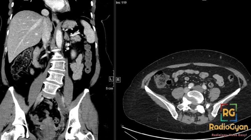
Varicocele. Left renal vein compression of more than 50 percent was present on contrast CT in 78 percent of men with a left varicocele against 10 percent of matched controls [23], so look at the vein in every left varicocele. In adolescents, however, finding the nutcracker phenomenon did not change testicular volume discrepancy or the rate of surgery [13].
Treatment and post-treatment imaging
Management runs from observation to open surgery, and most of the evidence is small retrospective series. Conservative management comes first, especially in children, where 86.8 percent of 423 reported patients were treated conservatively and 94.9 percent resolved or improved [4]. For interventional radiology training and fellowship pathways see the interventional radiology guide.
| Treatment | Symptom resolution | Reintervention | Imaging notes |
|---|---|---|---|
| Conservative (observation, weight gain) | 52 percent [24] | Not applicable | Duplex follow-up; spontaneous resolution common in children |
| Left renal vein transposition (to a lower point on the IVC) | 92 percent [24]; 87 percent in a 37-patient Mayo series [25] | 28.5 percent, the highest of any option [24]; freedom from reintervention 68 percent at 24 months [25] | Look for anastomotic restenosis; secondary stenting is the usual rescue |
| Endovascular left renal vein stenting | 76 percent [24] | 11.3 percent [24] | Check position and patency; migration to IVC, right atrium or ventricle |
| Laparoscopic or robotic extravascular stenting | 80 percent [24] | None reported [24] | Aortomesenteric angle rose from 20.6 to 44.5 degrees after placement [24] |
| Left gonadal vein transposition | 61 percent [24] | None reported [24] | Accepted by the Delphi panel as an alternative in selected patients [2] |
| Renal autotransplantation | 69 percent [24] | 7.2 percent [24] | Kidney in the iliac fossa; no Delphi consensus on effectiveness [2] |
The stent debate. In a 75-patient series followed for a mean 55 months, stents migrated in 5 (6.7 percent), all men: two into the inferior vena cava, one into the right atrium, one into the right ventricle and one along the renal vein [26]. That is the evidence behind the Delphi statement against primary stenting [2]. The counterweight is a 2026 series of 43 patients with a median follow-up of 46.5 months, dedicated venous stents in later cases and IVUS guidance in 74.4 percent, with no migration, primary patency of 89.9 percent at 1 year and 82.8 percent at 4 years, and symptom resolution in 90.7 percent [27]. Pooled data give similar haematuria resolution for endovascular and extravascular stenting, 86 and 89 percent [28].
What you will be asked after treatment. The panel agreed on first imaging, CT or duplex, within 6 weeks of surgery, yearly duplex in long-term follow-up, and specific assessment of stent patency or the degree of renal vein stenosis [2]. On a stent follow-up, confirm the stent is still spanning the aortomesenteric segment and has not moved into the cava, measure in-stent velocity against the hilar segment, and look again at the gonadal vein and pelvic varices. The panel also agreed that if a technically successful transposition does not relieve symptoms, the diagnosis should be reconsidered.
How to report a suspected nutcracker
- State the anatomy: anterior (aortomesenteric) or posterior (retroaortic vein), and any circumaortic or retroaortic variant [9].
- Give the measurements: hilar and aortomesenteric left renal vein diameters with the ratio, the aortomesenteric angle on a sagittal reformat, and whether a beak sign is present [5].
- On Doppler, give peak velocities at both points with the ratio, supine and upright if performed [15].
- Describe collaterals: left gonadal vein calibre and reflux, pelvic varices, ascending lumbar and hemiazygos collaterals, renal hilar varices. A refluxing collateral is what separates syndrome from phenomenon [19].
- Exclude the alternatives: renal vein thrombus, renal mass, stones, urothelial lesion, duodenal distension of superior mesenteric artery syndrome.
- Word the conclusion carefully: without the clinical picture, write left renal vein compression (nutcracker anatomy) and suggest clinical correlation, not nutcracker syndrome [1].
- If the patient is being considered for intervention, suggest venography with renocaval pressure measurement, with or without IVUS [3].
Frequently asked questions
References
- Ribeiro FS, Puech-Leão P, Zerati AE, Nahas WC, David-Neto E, De Luccia N. Prevalence of left renal vein compression (nutcracker phenomenon) signs on computed tomography angiography of healthy individuals. J Vasc Surg Venous Lymphat Disord. 2020;8(6):1058-1065. PMID 32335330.
- Heilijgers F, Gloviczki P, O’Sullivan G, Chavent B, Avgerinos ED, Harth K, et al. Nutcracker syndrome (a Delphi consensus). J Vasc Surg Venous Lymphat Disord. 2025;13(1):101970. PMID 39362632.
- Nastasi DR, Fraser AR, Williams AB, Bhamidi V. A systematic review on nutcracker syndrome and proposed diagnostic algorithm. J Vasc Surg Venous Lymphat Disord. 2022;10(6):1410-1416. PMID 36007798.
- Meyer J, Rother U, Stehr M, Meyer A. Nutcracker syndrome in children: appearance, diagnostics, and treatment – a systematic review. J Pediatr Surg. 2022;57(11):716-722. PMID 35065803.
- Kim KW, Cho JY, Kim SH, Yoon JH, Kim DS, Chung JW, et al. Diagnostic value of computed tomographic findings of nutcracker syndrome: correlation with renal venography and renocaval pressure gradients. Eur J Radiol. 2011;80(3):648-654. PMID 20869828.
- Qin Y, Tian L, Chen X, Li Q. The superior mesenteric artery angle in diagnosis of nutcracker syndrome: a systematic review and meta-analysis. Abdom Radiol (NY). 2025;50(2):851-859. PMID 39177776.
- Kim SH, Cho SW, Kim HD, Chung JW, Park JH, Han MC. Nutcracker syndrome: diagnosis with Doppler US. Radiology. 1996;198(1):93-97. PMID 8539413.
- Kubiszewski K, Clingan J, Caserta MP. Sonographic hallmarks of nutcracker syndrome. Ultrasound Q. 2026;42(1):e00734. PMID 41705917.
- Heidler S, Hruby S, Schwarz S, Sellner-Zwieauer Y, Hoeltl W, Albrecht W. Prevalence and incidence of clinical symptoms of the retroaortic left renal vein. Urol Int. 2015;94(2):173-176. PMID 25661199.
- Grimm LJ, Engstrom BI, Nelson RC, Kim CY. Incidental detection of nutcracker phenomenon on multidetector CT in an asymptomatic population: prevalence and associated findings. J Comput Assist Tomogr. 2013;37(3):415-418. PMID 23674014.
- Park JH, Lee GH, Lee SM, Eisenhut M, Kronbichler A, Lee KH, et al. Posterior nutcracker syndrome – a systematic review. Vasa. 2018;47(1):23-29. PMID 29165061.
- Mazzoni MB, Kottanatu L, Simonetti GD, Ragazzi M, Bianchetti MG, Fossali EF, et al. Renal vein obstruction and orthostatic proteinuria: a review. Nephrol Dial Transplant. 2011;26(2):562-565. PMID 20656752.
- Hannick JH, Blais AS, Kim JK, Traubici J, Shiff M, Book R, et al. Prevalence, Doppler ultrasound findings, and clinical implications of the nutcracker phenomenon in pediatric varicoceles. Urology. 2019;128:78-83. PMID 30885542.
- Kim SH. Doppler US and CT diagnosis of nutcracker syndrome. Korean J Radiol. 2019;20(12):1627-1637. PMID 31854150.
- Romera-Villegas A, Vila-Coll R, Cairols-Castellote M, Poveda-Monge R, Masuet-Aumatell C, Grinyó-Boira M. The importance of a standing position in the diagnosis of nutcracker phenomenon by duplex sonography. Int Angiol. 2009;28(6):461-468. PMID 20087283.
- Cheon JE, Kim WS, Kim IO, Kim SH, Yeon KM, Ha IS, et al. Nutcracker syndrome in children with gross haematuria: Doppler sonographic evaluation of the left renal vein. Pediatr Radiol. 2006;36(7):682-686. PMID 16770671.
- Park SJ, Lim JW, Cho BS, Yoon TY, Oh JH. Nutcracker syndrome in children with orthostatic proteinuria: diagnosis on the basis of Doppler sonography. J Ultrasound Med. 2002;21(1):39-45. PMID 11794401.
- Takebayashi S, Ueki T, Ikeda N, Fujikawa A. Diagnosis of the nutcracker syndrome with color Doppler sonography: correlation with flow patterns on retrograde left renal venography. AJR Am J Roentgenol. 1999;172(1):39-43. PMID 9888735.
- Yun SJ, Lee JM, Nam DH, Ryu JK, Lee SH. Discriminating renal nutcracker syndrome from asymptomatic nutcracker phenomenon using multidetector computed tomography. Abdom Radiol (NY). 2016;41(8):1580-1588. PMID 27221972.
- Arthurs OJ, Mehta U, Set PA. Nutcracker and SMA syndromes: what is the normal SMA angle in children? Eur J Radiol. 2012;81(8):e854-e861. PMID 22579528.
- Atasoy D, Cansu A, Bekirçavuşoğlu AF, Özdoğan EB, Ahmetoğlu A. The utility of magnetic resonance angiography in children with nutcracker syndrome. Turk J Med Sci. 2021;51(5):2396-2402. PMID 33992039.
- Meissner MH, Khilnani NM, Labropoulos N, Gasparis AP, Gibson K, Greiner M, et al. The Symptoms-Varices-Pathophysiology classification of pelvic venous disorders: a report of the American Vein and Lymphatic Society International Working Group on Pelvic Venous Disorders. J Vasc Surg Venous Lymphat Disord. 2021;9(3):568-584. PMID 33529720.
- Lewis DS, Grimm LJ, Kim CY. Left renal vein compression as cause for varicocele: prevalence and associated findings on contrast-enhanced CT. Abdom Imaging. 2015;40(8):3147-3151. PMID 26210372.
- Sarikaya S, Altas O, Ozgur MM, Hancer H, Aksut M, Topcu KO, et al. Contemporary management of nutcracker syndrome: a systematic review. Ann Vasc Surg. 2025;121:406-421. PMID 40816484.
- Erben Y, Gloviczki P, Kalra M, Bjarnason H, Reed NR, Duncan AA, et al. Treatment of nutcracker syndrome with open and endovascular interventions. J Vasc Surg Venous Lymphat Disord. 2015;3(4):389-396. PMID 26992616.
- Wu Z, Zheng X, He Y, Fang X, Li D, Tian L, et al. Stent migration after endovascular stenting in patients with nutcracker syndrome. J Vasc Surg Venous Lymphat Disord. 2016;4(2):193-199. PMID 26993867.
- Anan H, Sudina Y, Sridharan N, Hager E, Eid R, Avgerinos E, et al. Stenting for symptomatic left renal vein compression in patients with complex pelvic venous disease is safe on long-term follow-up. J Vasc Surg Venous Lymphat Disord. 2026;14(4):102469. PMID 41780673.
- Fuentes-Perez A, Bush RL, Kalra M, Shortell C, Gloviczki P, Brigham TJ, et al. Systematic review of endovascular versus laparoscopic extravascular stenting for treatment of nutcracker syndrome. J Vasc Surg Venous Lymphat Disord. 2023;11(2):433-441. PMID 36404475.

Improve Content And Help Your Colleagues!
Found an error in the post? Please let us know using our contact page and we will update it with due credits!
If you wish to contribute radiological images for this case (or any other article on the website), you can submit them here and you will be duly credited: Submit radiology case