


- Splenic index (L×W×T): 120–480
- Splenic volume: 107–315 cm³ (mean ~215 cm³)
- Splenic weight: under 230 g
What this splenic index calculator does
Enter the three orthogonal spleen dimensions (length, width and thickness in cm) measured on CT, MRI or ultrasound and the tool instantly returns the splenic index, estimated splenic volume and weight, and flags whether the spleen is normal or enlarged. Use the Copy report button to paste a ready-made sentence into your report.
Formulae used
- Splenic index = Length × Width × Thickness (the product of the three dimensions in cm).
- Splenic volume (cm³) = 30 + 0.58 × (Length × Width × Thickness) — the regression equation of Prassopoulos et al.
- Splenic weight (g) = Volume × 1.05, assuming a splenic tissue density of ~1.05 g/cm³.
How to measure length, width and thickness
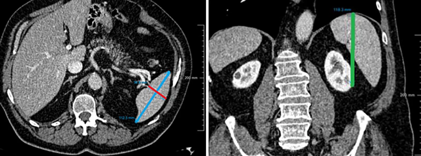
- Length (L): cranio-caudal distance between the first and last slices on which the spleen is seen in the axial plane.
- Width (W): largest measurable long-axis diameter in the axial plane.
- Thickness (T): largest dimension perpendicular to the width in the axial plane.
Click here for an illustration on CT. The same three measurements are used whether they come from CT, MRI or ultrasound.
Normal reference values
| Parameter | Normal adult range |
|---|---|
| Splenic index (L×W×T) | 120–480 |
| Splenic volume | 107–315 cm³ (mean ~215 cm³) |
| Splenic weight | Under 230 g |
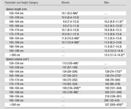
Normal splenic size varies with body height and sex — taller people and men tend to have larger spleens. The chart below (from Chow et al., Radiology 2016) shows how normal spleen size changes with age, sex and height.
Interpreting the result
A volume above ~315 cm³ (or index above 480) indicates splenomegaly. Common causes include portal hypertension and cirrhosis, haematological malignancy (lymphoma, leukaemia, myeloproliferative disease), and infection (mononucleosis, malaria). A spleen below the normal range may be seen in sickle-cell disease, coeliac disease and normal ageing. Always interpret the number alongside the clinical picture rather than in isolation.
Limitations
The volume figure is an estimate from a linear regression, not a true voxel-based segmentation, so it will differ slightly from software volumetry. Reference ranges are derived from adult cohorts and do not account for paediatric age, extremes of body size or ethnicity. Use it as a fast bedside estimate, not a definitive measurement.
References
- Chow KU, Luxembourg B, Seifried E, Bonig H. Spleen Size Is Significantly Influenced by Body Height and Sex: Establishment of Normal Values for Spleen Size at US with a Cohort of 1200 Healthy Individuals. Radiology. 2016;279(1):306-313. doi:10.1148/radiol.2015150887
- Prassopoulos P, Daskalogiannaki M, Raissaki M, Hatjidakis A, Gourtsoyiannis N. Determination of normal splenic volume on computed tomography in relation to age, gender and body habitus. Eur Radiol. 1997;7(2):246-248. doi:10.1007/s003300050145
- Gilbert T, Castellino RA. The spleen in Hodgkin disease: diagnostic value of CT. Invest Radiol. 1986;21(5):437-439. doi:10.1097/00004424-198605000-00017
Check out other radiology calculators:
Disclaimer: This tool is for educational purposes only and is not a substitute for clinical judgment.

Improve Content And Help Your Colleagues!
Found an error in the post? Please let us know using our contact page and we will update it with due credits!
If you wish to contribute radiological images for this case (or any other article on the website), you can submit them here and you will be duly credited: Submit radiology case
About the Author
Dr. Amar Udare, MD, DNB
Dr. Udare holds an MBBS and MD degree, and his expertise lies in the field of radiology. He has authored multiple peer-reviewed publications, contributing significantly to the medical field. His works can be accessed on PubMed and Google Scholar.
In addition to his academic and professional achievements, Dr. Udare is an avid reader and enjoys exploring the latest advancements in medical technology. His commitment to making complex medical knowledge accessible to patients and the general public aligns with our mission at RadioGyan.com.
For any further questions or clarifications, feel free to reach out to Dr. Udare via the contact form.