


How to Measure Prostate Volume
Normal Prostate Volume and PSA (Age Specific)
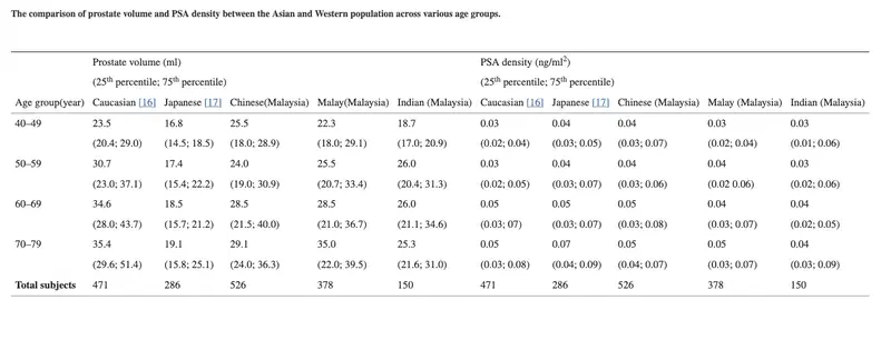
PSA density cut off used is 0.15.
Prostate Volume Calculator: Normal Prostate Volume by Age and PSA Density
Key facts for practice and radiology board exams
Normal prostate volume is about 25 to 30 mL in men under 50 and grows roughly 0.6 mL a year after that, so 35 to 40 mL is unremarkable at 70 [1]. Volume = AP × TR × CC × 0.52. PSA density = serum PSA ÷ volume; after a negative MRI, a density below 0.10 ng/mL/cc leaves roughly a 4% chance of clinically significant cancer [2].
- Measure AP and CC on the midsagittal image, TR on axial, then multiply by 0.52 (the prolate ellipsoid coefficient, written 0.523 in some sources). This is the PI-RADS v2.1 method [3].
- Over 30 mL is prostatomegaly: 30 to 50 mL mild, 50 to 80 mL moderate, over 80 mL marked. Read it against the patient’s age before calling it abnormal.
- 0.15 ng/mL/cc is the conventional PSA density alarm level, not a rule-out. Used alone in the pre-MRI era it missed about half of all cancers [4].
- PSA density earns its place after the MRI, not before it. With a negative scan (PI-RADS 1 to 2), the post-test probability of significant cancer is about 6% below 0.15 and 4% below 0.10 [2].
- 5-alpha-reductase inhibitors roughly halve PSA. Double the measured value before calculating density in a man on finasteride or dutasteride.
What is a normal prostate volume by age?
Normal prostate volume is 25 to 30 mL in middle age and climbs steadily thereafter. In the Baltimore Longitudinal Study of Aging the median was 28 mL at a median age of 58, growing about 0.6 mL per year [1]. Growth is not uniform: a minority of men account for most of the increase, which is why age-matched tables are a guide rather than a threshold.
| Age group | Caucasian | Chinese (Malaysia) | Malay (Malaysia) | Indian (Malaysia) |
|---|---|---|---|---|
| 40 to 49 | 23.5 | 25.5 | 22.3 | 18.7 |
| 50 to 59 | 30.7 | 24.0 | 25.5 | 26.0 |
| 60 to 69 | 34.6 | 28.5 | 28.5 | 26.0 |
| 70 to 79 | 35.4 | 29.1 | 35.0 | 25.3 |
The same study found that PSA and PSA density differ by ethnic group at the same age, with Indian men having the lowest median PSA and prostate volume [5]. A single threshold is therefore not equally calibrated in every population.
| Volume | Descriptor |
|---|---|
| Up to 30 mL | Normal |
| 30 to 50 mL | Mild prostatomegaly |
| 50 to 80 mL | Moderate prostatomegaly |
| Over 80 mL | Marked prostatomegaly |
How to measure prostate volume on MRI and ultrasound
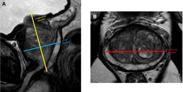
Measure the anteroposterior and craniocaudal dimensions on the midsagittal T2 image and the transverse dimension on an axial image, then apply the prolate ellipsoid formula. The 0.52 coefficient is pi divided by 6. This is the method specified in the PI-RADS v2.1 update [3].
- Volume (mL) = AP × TR × CC × 0.52
- PSA density (ng/mL/cc) = serum PSA ÷ prostate volume
Method matters more than the decimal places. Comparing contemporary techniques against manual 3D segmentation on MRI, the MRI ellipsoid formula agreed closely (ICC 0.90) while the ultrasound ellipsoid volume agreed only moderately (ICC 0.73), and formula-based volumes scattered more widely than segmentation-based ones [7]. A TRUS volume and an MRI volume are not interchangeable in the denominator of a PSA density.
PSA density: thresholds and what the evidence actually shows
PSA density normalises serum PSA to gland size, correcting for the PSA that a large benign prostate produces on its own. The conventional threshold is 0.15 ng/mL/cc. The table below is the honest version of the evidence: the cut-off behaves very differently before and after an MRI, and the pre-MRI studies are far less reassuring than the number’s popularity suggests.
| PSA density | Setting | What the study found | Source |
|---|---|---|---|
| 0.15 | PSA 4 to 10, no MRI | Improved specificity but missed about half of all cancers, including 47% of organ-confined cancers that a PSA above 4 detected. The authors recommended biopsying on PSA rather than PSA density. | Catalona 1994 [4] |
| 0.07 or below | Biopsy decision, systematic biopsy era | Would have avoided 19.7% of biopsies while missing 6.9% of Gleason 7 or higher cancers. | Nordström 2018 [8] |
| Below 0.09 | Men referred for biopsy | 4% chance of clinically significant cancer. The Youden-optimal cut-off in the same cohort was 0.20 (70% sensitivity, 79% specificity). | Yusim 2020 [9] |
| 0.10 / 0.15 | Biopsy-naive / prior negative biopsy | Optimal cut-offs for separating cancer from intraprostatic inflammation as the cause of a raised PSA. | Bruno 2021 [10] |
| Below 0.15 | Negative MRI (PI-RADS 1 to 2) | Post-test probability of clinically significant cancer 6%. | Wang 2024 [2] |
| Below 0.10 | Negative MRI (PI-RADS 1 to 2) | Post-test probability of clinically significant cancer 4%. | Wang 2024 [2] |
Where PSA density changes management
- Negative MRI. This is the setting where PSA density is most useful. Pooled across 39 studies, a PI-RADS 1 to 2 scan plus a density below 0.10 leaves a 4% residual risk of significant cancer, low enough that many pathways defer biopsy [2].
- PI-RADS 3 lesions. PSA density is the usual tiebreaker for an equivocal scan. Treat it as a probability modifier shifting an already borderline decision, not as a threshold that settles it.
- Active surveillance. A density below 0.15 remains a long-standing inclusion criterion for low-risk disease.
- After a benign systematic biopsy. In a screening cohort followed for a median of 17 years, 15-year prostate cancer mortality was 0.5% with a density below 0.15 against 2.0% at or above it [11], so the value at the time of a negative biopsy carries long-term prognostic weight.
Pitfalls that invalidate a PSA density
- 5-alpha-reductase inhibitors. Finasteride and dutasteride roughly halve serum PSA. Double the measured PSA before calculating density.
- Prostatitis. Inflammation raises PSA out of proportion to volume and inflates the density. PSA density separates cancer from inflammation better than PSA alone, but it does not abolish the problem [10].
- Mixing modalities. An ultrasound ellipsoid volume and an MRI ellipsoid volume are not the same number [7]. State which one you used in the report.
- Using transition zone volume. The denominator is whole gland volume. Substituting the transition zone inflates the density and is not what the thresholds were derived from.
- Treating the cut-off as a diagnosis. Every threshold above is a shift in probability. None of them is sensitive enough to rule cancer out on its own.
Frequently asked questions
References
- Loeb S, Kettermann A, Carter HB, Ferrucci L, Metter EJ, Walsh PC. Prostate volume changes over time: results from the Baltimore Longitudinal Study of Aging. J Urol. 2009;182(4):1458-62.
- Wang S, Kozarek J, Russell R, Drescher M, Khan A, Kundra V, et al. Diagnostic performance of prostate-specific antigen density for detecting clinically significant prostate cancer in the era of magnetic resonance imaging: a systematic review and meta-analysis. Eur Urol Oncol. 2024;7(2):189-203.
- Turkbey B, Rosenkrantz AB, Haider MA, Padhani AR, Villeirs G, Macura KJ, et al. Prostate Imaging Reporting and Data System version 2.1: 2019 update of Prostate Imaging Reporting and Data System version 2. Eur Urol. 2019;76(3):340-51.
- Catalona WJ, Richie JP, deKernion JB, Ahmann FR, Ratliff TL, Dalkin BL, et al. Comparison of prostate specific antigen concentration versus prostate specific antigen density in the early detection of prostate cancer: receiver operating characteristic curves. J Urol. 1994;152(6 Pt 1):2031-6.
- Lim J, Bhoo-Pathy N, Sothilingam S, Malek R, Sundram M, Hisham Bahadzor B, et al. Ethnicity is an independent determinant of age-specific PSA level: findings from a multiethnic Asian setting. PLoS One. 2014;9(8):e104917.
- Oesterling JE, Jacobsen SJ, Chute CG, Guess HA, Girman CJ, Panser LA, et al. Serum prostate-specific antigen in a community-based population of healthy men: establishment of age-specific reference ranges. JAMA. 1993;270(7):860-4.
- Bezinque A, Moriarity A, Farrell C, Peabody H, Noyes SL, Lane BR. Determination of prostate volume: a comparison of contemporary methods. Acad Radiol. 2018;25(12):1582-7.
- Nordström T, Akre O, Aly M, Grönberg H, Eklund M. Prostate-specific antigen (PSA) density in the diagnostic algorithm of prostate cancer. Prostate Cancer Prostatic Dis. 2018;21(1):57-63.
- Yusim I, Krenawi M, Mazor E, Novack V, Mabjeesh NJ. The use of prostate specific antigen density to predict clinically significant prostate cancer. Sci Rep. 2020;10(1):20015.
- Bruno SM, Falagario UG, d’Altilia N, Recchia M, Mancini V, Selvaggio O, et al. PSA density help to identify patients with elevated PSA due to prostate cancer rather than intraprostatic inflammation: a prospective single center study. Front Oncol. 2021;11:693684.
- Pylväläinen J, Talala K, Raitanen J, Rannikko A, Auvinen A. Association of prostate-specific antigen density with prostate cancer mortality after a benign systematic prostate biopsy result. BJU Int. 2025;135(5):841-50.
Related: PI-RADS calculator. Browse all radiology calculators.
This calculator is for educational use. Where a local protocol or the current ACR and EAU source documents differ from the thresholds quoted here, those take precedence.

Improve Content And Help Your Colleagues!
Found an error in the post? Please let us know using our contact page and we will update it with due credits!
If you wish to contribute radiological images for this case (or any other article on the website), you can submit them here and you will be duly credited: Submit radiology case
About the Author
Dr. Amar Udare, MD, DNB
Dr. Udare holds an MBBS and MD degree, and his expertise lies in the field of radiology. He has authored multiple peer-reviewed publications, contributing significantly to the medical field. His works can be accessed on PubMed and Google Scholar.
In addition to his academic and professional achievements, Dr. Udare is an avid reader and enjoys exploring the latest advancements in medical technology. His commitment to making complex medical knowledge accessible to patients and the general public aligns with our mission at RadioGyan.com.
For any further questions or clarifications, feel free to reach out to Dr. Udare via the contact form.