


| Cat. | Descriptor | Key findings | Management | Malignancy |
|---|---|---|---|---|
| 0 | Incomplete | Part/all of lungs not evaluable, awaiting prior CT, or suspected infection/inflammation | Compare to prior CT, additional imaging, and/or 1–3 mo LDCT | n/a |
| 1 | Negative | No nodules, or nodule with benign (complete/central/popcorn/ring) calcification or fat | Annual LDCT (12 mo) | < 1% |
| 2 | Benign | Solid < 6 mm (new < 4 mm); part-solid < 6 mm total; GGN < 30 mm; juxtapleural ≤ 10 mm | Annual LDCT (12 mo) | < 1% |
| 3 | Probably benign | Solid ≥ 6–< 8 mm (new 4–< 6); part-solid ≥ 6 mm total w/ solid < 6 mm; GGN ≥ 30 mm | 6-month LDCT | 1–2% |
| 4A | Suspicious | Solid ≥ 8–< 15 mm (new 6–< 8); part-solid solid comp. ≥ 6–< 8 mm; segmental airway (baseline) | 3-month LDCT; PET/CT if ≥ 8 mm solid | 5–15% |
| 4B | Very suspicious | Solid ≥ 15 mm (new/growing ≥ 8); part-solid solid comp. ≥ 8 mm; slow-growth over serial exams | Diagnostic CT, PET/CT, ± tissue sampling | > 15% |
| 4X | Very suspicious + | Category 3/4 nodule with extra features increasing suspicion (spiculation, nodal/metastatic disease, GGN doubling) | As for 4B | > 15% |
| S | Modifier | Significant/potentially significant finding unrelated to lung cancer (added to category 0–4) | Per ACR Incidental Findings | — |
This tool implements ACR Lung-RADS® v2022 rules. Final classification and management must be made by a qualified radiologist using the full imaging study and clinical context.
Lung-RADS® v2022 Calculator for Low-Dose CT Lung Cancer Screening
This Lung-RADS calculator assigns the correct Lung Imaging Reporting and Data System version 2022 assessment category to a pulmonary nodule on a low-dose CT (LDCT) lung cancer screening study, and generates an editable, dictation-ready report. Enter the nodule type, its relationship to prior exams, and its mean diameter (plus the solid-component size for part-solid nodules), and the tool returns the Lung-RADS category, the approximate probability of malignancy, and the ACR-recommended management.
Version 2022, released in November 2022, is the current edition and replaces v1.1. It adds explicit rules for atypical pulmonary cysts, juxtapleural nodules and airway-centred nodules, clarifies the growth definition, and introduces stepped management for slowly growing lesions. The calculator implements these rules and produces report text you can paste into PowerScribe, RadAI or any reporting system.
When Lung-RADS Applies
Lung-RADS is intended for asymptomatic screening of patients who meet lung cancer screening eligibility criteria, imaged with a dedicated LDCT protocol. It is not designed for diagnostic chest CT, for the work-up of a symptomatic patient, or for patients with a known active malignancy under surveillance. Each examination is coded by its most suspicious nodule; the category and management recommendation that follow apply to the whole study, not to every nodule individually.
How to Use This Lung-RADS Calculator
- Select the dominant finding – the nodule with the highest degree of suspicion, since each exam is coded by its most concerning nodule.
- Choose the timepoint – baseline screen, a new nodule, a growing nodule (> 1.5 mm increase within 12 months), or a stable/slowly growing nodule.
- Enter the mean diameter – the average of the long and short axis to one decimal point on lung windows. For part-solid nodules, also enter the solid-component diameter.
- Add any modifiers – features that increase suspicion for lung cancer (category 4X) or a clinically significant non-lung finding (S modifier).
The category, approximate malignancy risk and management recommendation update automatically as you complete each field, and a copyable report block appears for any classifiable finding.
Categories, Malignancy Risk and Management
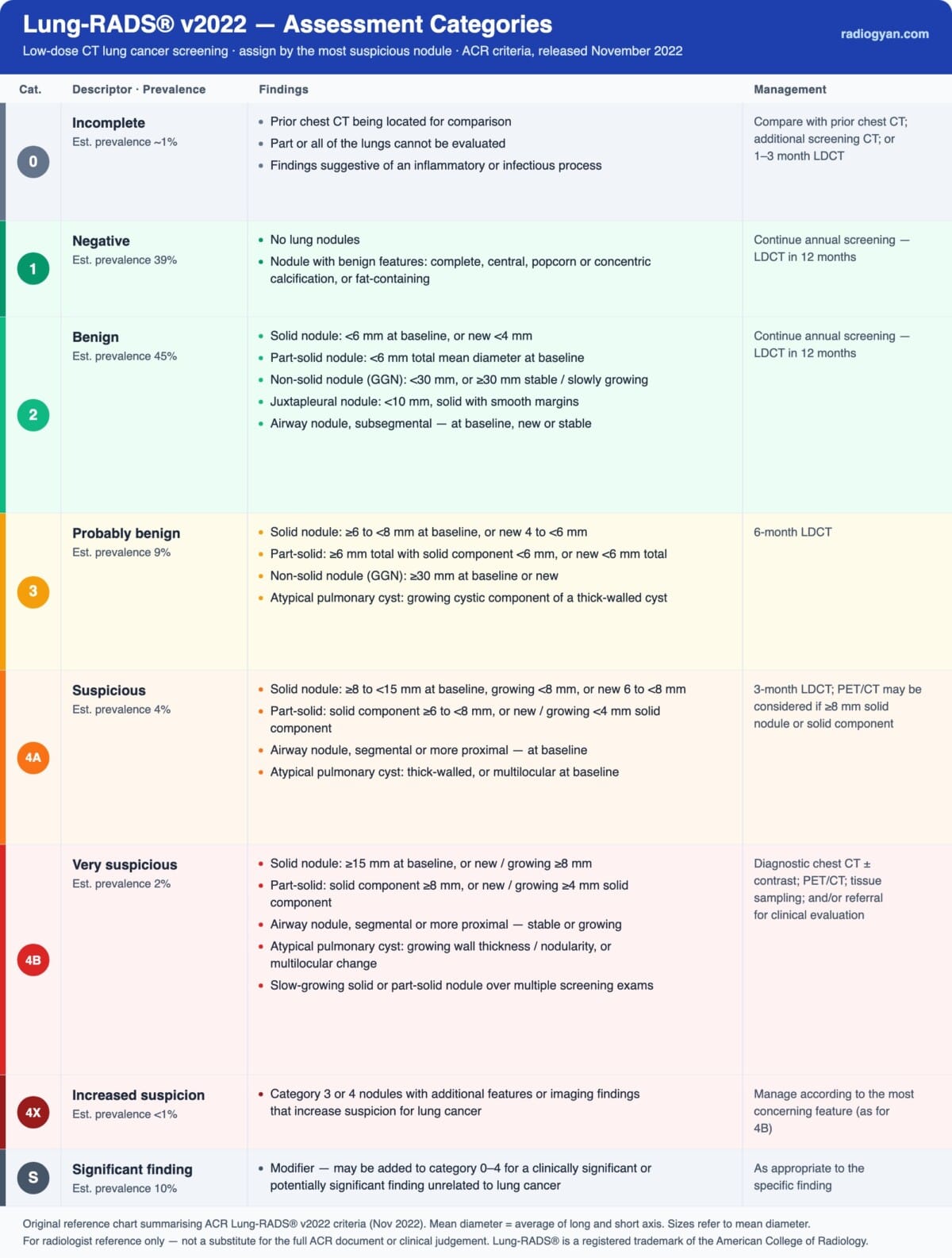
Link to original ACR reference chart: Lung-RADS 2022 ACR Reference Chart
| Category | Meaning | Approx. malignancy | Management |
|---|---|---|---|
| 0 | Incomplete | n/a | Compare with prior CT, additional imaging, and/or 1–3 month LDCT |
| 1 | Negative | < 1% | Annual LDCT (12 months) |
| 2 | Benign | < 1% | Annual LDCT (12 months) |
| 3 | Probably benign | 1–2% | 6-month LDCT |
| 4A | Suspicious | 5–15% | 3-month LDCT; PET/CT if ≥ 8 mm solid component |
| 4B | Very suspicious | > 15% | Diagnostic chest CT, PET/CT, ± tissue sampling |
| 4X | Category 3 or 4 with additional suspicious features | > 15% | As for 4B; individualise to the additional feature |
Size Thresholds by Nodule Type
Solid nodules
- Category 2: < 6 mm at baseline, or new < 4 mm.
- Category 3: ≥ 6 to < 8 mm at baseline, or new 4 to < 6 mm.
- Category 4A: ≥ 8 to < 15 mm at baseline, new 6 to < 8 mm, or growing < 8 mm.
- Category 4B: ≥ 15 mm at baseline, or new/growing ≥ 8 mm.
Part-solid nodules
- Category 2: < 6 mm total mean diameter at baseline.
- Category 3: ≥ 6 mm total with solid component < 6 mm at baseline, or new < 6 mm total.
- Category 4A: solid component ≥ 6 to < 8 mm at baseline, or new/growing solid component < 4 mm.
- Category 4B: solid component ≥ 8 mm at baseline, or new/growing solid component ≥ 4 mm.
Non-solid (ground-glass) nodules
- Category 2: < 30 mm at baseline, new, or growing; or ≥ 30 mm that is stable or slowly growing.
- Category 3: ≥ 30 mm at baseline or new.
How to Measure a Nodule for Lung-RADS
Measure both the long and short axis to one decimal point in millimetres and report the mean diameter — their average — to one decimal point. The axes may be obtained in any plane to reflect the true size of the nodule, and need not lie within a single image. For part-solid nodules, measure both the total nodule and the solid component, because the solid component drives the category from 3 upward. When volumetry is used, report volumes to the nearest whole cubic millimetre. Measure on lung windows, and exclude adjacent vessels, atelectasis and surrounding ground-glass from the solid measurement.
Growth, New Nodules and Stepped Management
Growth is defined as an increase in mean diameter of more than 1.5 mm within a 12-month interval. A nodule that crosses into a higher size category should be re-classified by size even when it does not formally meet the growth definition. A key v2022 change is the recognition of slow growth: a solid or part-solid nodule that enlarges over multiple screening rounds without crossing the 1.5 mm/12-month threshold is nonetheless suspicious and is classified as category 4B. Conversely, v2022 introduces stepped management — a category 3 or 4A nodule that is stable or decreasing at follow-up can be stepped down toward annual screening rather than held indefinitely at short interval.
Special Categories in v2022
- Juxtapleural nodules: solid, smoothly marginated, oval, lentiform or triangular nodules < 10 mm are category 2 (typical intrapulmonary lymph nodes).
- Atypical pulmonary cysts: thin-walled, uniform cysts (wall < 2 mm) are benign and not classified within Lung-RADS, but a thick or asymmetric wall, a multilocular cyst, or an enlarging or growing solid/soft-tissue component raises the category up to 4A or 4B.
- Airway (endobronchial) nodules: a segmental or more proximal airway nodule is category 4A at baseline; if it resolves or clears on a 3-month CT it is category 1, whereas a nodule that is stable or growing on follow-up is category 4B.
- Suspected infection or inflammation: findings likely to be infectious or inflammatory may be assigned category 0 for short-interval (1–3 month) LDCT and re-classified once resolution or persistence is established.
The S and 4X Modifiers
Category 4X applies to a category 3 or 4 nodule with additional imaging features that increase suspicion for lung cancer — spiculation, lymphadenopathy, frank metastatic disease, or a ground-glass nodule that doubles in size within one year. The S modifier can be appended to any category (0–4) to flag a clinically significant or potentially significant finding unrelated to lung cancer, managed according to the ACR Incidental Findings recommendations. The two are independent: a study can be, for example, 4X for a spiculated nodule and carry an S modifier for a separate adrenal mass.
What’s New in Lung-RADS v2022
- Ground-glass nodules: non-solid nodules < 30 mm are now category 2, reducing unnecessary follow-up.
- Slow growth: a solid or part-solid nodule growing over serial exams without meeting the 1.5 mm/12-month threshold is category 4B.
- Atypical pulmonary cysts and airway (endobronchial) nodules have explicit classification and follow-up criteria.
- Juxtapleural nodules < 10 mm with benign morphology are formally category 2.
- Stepped management allows down-classification of stable or decreasing category 3/4A nodules, and the growth definition is clarified as > 1.5 mm in mean diameter within 12 months.
Reporting Tips and Common Pitfalls
- Code the exam by its most suspicious nodule; do not average or downplay a dominant finding because most nodules are benign.
- For part-solid nodules, the solid component — not the total diameter — drives categories 3 and above; measure and report it explicitly.
- Apply the new-nodule thresholds (lower than baseline) when a nodule was not present on a prior screen, and re-classify by size whenever a nodule crosses a threshold even without meeting the growth definition.
- Do not forget the S modifier for significant extrapulmonary findings; it does not change the lung category but must be communicated.
- Follow-up timing is counted from the date of the exam being interpreted, not from the prior study or the report date.
Frequently Asked Questions
What does a positive Lung-RADS screen mean?
A positive screen is Lung-RADS category 3 or 4; categories 1 and 2 are negative. The split defines a programme’s positive-screen and recall rates, and only category 3–4 studies trigger short-interval or diagnostic follow-up.
How is Lung-RADS growth defined?
Growth is an increase in mean diameter of more than 1.5 mm within a 12-month interval. A nodule that crosses into a higher size category should be re-classified by size even if it does not formally meet the growth definition, and a solid or part-solid nodule that grows slowly over serial exams is category 4B.
When does follow-up timing start?
The timing of follow-up imaging is measured from the date of the exam being interpreted — for example, a category 2 annual-LDCT recommendation is counted 12 months from the current exam.
What is Lung-RADS category 4X?
4X is a category 3 or 4 nodule with additional features that raise suspicion for malignancy — spiculation, lymphadenopathy, frank metastatic disease, or a ground-glass nodule that doubles in size within one year — and is managed as 4B (> 15% malignancy risk). It is independent of the S modifier, which flags a clinically significant non-lung finding on the same study.
References
- American College of Radiology. Lung-RADS® – Lung Imaging Reporting and Data System. Reston, VA: ACR (Lung-RADS v2022 release notes and assessment categories).
- Christensen J, Prosper AE, Wu CC, et al. ACR Lung-RADS v2022: Assessment Categories and Management Recommendations. Journal of the American College of Radiology. 2024;21(3):473–488.
- The National Lung Screening Trial Research Team. Reduced Lung-Cancer Mortality with Low-Dose Computed Tomographic Screening. New England Journal of Medicine. 2011;365(5):395–409.
- Pinsky PF, Gierada DS, Black W, et al. Performance of Lung-RADS in the National Lung Screening Trial: a retrospective assessment. Annals of Internal Medicine. 2015;162(7):485–491.
- US Preventive Services Task Force. Screening for Lung Cancer: US Preventive Services Task Force Recommendation Statement. JAMA. 2021;325(10):962–970.
This calculator implements Lung-RADS® v2022 as published by the ACR; in case of any discrepancy, the official ACR Lung-RADS documents take precedence.

Improve Content And Help Your Colleagues!
Found an error in the post? Please let us know using our contact page and we will update it with due credits!
If you wish to contribute radiological images for this case (or any other article on the website), you can submit them here and you will be duly credited: Submit radiology case
About the Author
Dr. Amar Udare, MD, DNB
Dr. Udare holds an MBBS and MD degree, and his expertise lies in the field of radiology. He has authored multiple peer-reviewed publications, contributing significantly to the medical field. His works can be accessed on PubMed and Google Scholar.
In addition to his academic and professional achievements, Dr. Udare is an avid reader and enjoys exploring the latest advancements in medical technology. His commitment to making complex medical knowledge accessible to patients and the general public aligns with our mission at RadioGyan.com.
For any further questions or clarifications, feel free to reach out to Dr. Udare via the contact form.