


45-year-old female presenting with headache. What is your diagnosis?
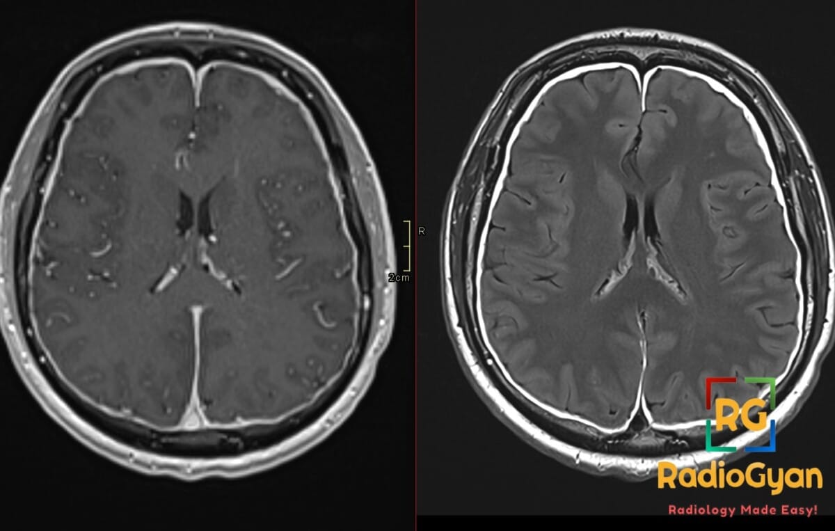
Diagnosis and teaching points:
Diagnosis: Subdural effusion and pachymeningeal enhancement in a patient with spontaneous intracranial hypotension.
Key facts for board exams:
- Diffuse smooth pachymeningeal enhancement after gadolinium is the hallmark MRI finding, sparing the leptomeninges.
- Bilateral subdural effusions are typically thin, convexity-based, and CSF-like in signal.
- Brain sag with low-lying cerebellar tonsils and reduced mamillopontine distance mimics Chiari I.
- CSF opening pressure is <60 mm H2O in only about one-third of patients; normal pressure does not exclude SIH.
- First-line treatment is lumbar epidural blood patch; targeted patching after leak localization if refractory.
Detailed teaching points:
- Clinical: Typically young to middle-aged adults; female predominance ~2:1; orthostatic headache that worsens on standing and improves with recumbency; associated tinnitus, aural pressure, nausea, and neck pain.
- Etiology/Pathophys: Spontaneous spinal CSF leak causes reduced CSF volume; compensatory venous hyperemia and brain sag produce the imaging findings; Monro-Kellie mechanism explains venous distension.
- CT: Noncontrast CT may show pseudo-subarachnoid-hemorrhage sign with hyperdense basal vessels against reduced CSF spaces; CT myelography can localize the leak, with dynamic CTM preferred for high-flow ventral tears.
- MRI: T1 post-contrast shows diffuse smooth pachymeningeal enhancement; T2 shows brainstem and tonsillar descent with narrowed cisterns; FLAIR highlights subdural effusions; GRE/SWI detects superficial siderosis or hemorrhage; spine MRI may show a T2-bright longitudinal epidural collection.
- Signs: SEEPS mnemonic – Subdural collections, Enhancement of pachymeninges, Engorgement of venous structures, Pituitary enlargement, Sagging of brain; venous distension sign – convex inferior border of transverse sinus; midbrain sagging sign – reduced mamillopontine distance; cortical vein sign – prominent veins traversing subdural collection; pseudo-SAH sign – hyperdense vessels on noncontrast CT.
- DDx: Postdural puncture headache – recent puncture or epidural history; Chiari I malformation – congenital tonsillar descent without pachymeningeal enhancement or venous engorgement; meningitis – fever, leptomeningeal involvement, irregular enhancement; idiopathic hypertrophic pachymeningitis – mass-like dural thickening rather than smooth enhancement; cerebral venous thrombosis – thrombus on venography, may complicate SIH.
- Tx: Conservative measures (bed rest, hydration, caffeine) for up to 1-2 weeks; lumbar epidural blood patch as first-line intervention, repeatable after 2-4 weeks; targeted blood or fibrin patch after CTM/DSM localization; surgery for refractory or life-threatening cases; follow-up brain MRI with contrast at 3-6 months to document resolution.
OSCE Questions
Question: What mnemonic summarizes the major intracranial MRI findings of this condition?
SEEPS: Subdural fluid collections, Enhancement of pachymeninges, Engorgement of venous structures, Pituitary enlargement, Sagging of the brain.
Question: What is the first-line imaging modality for this condition?
Contrast-enhanced brain MRI with whole-spine MRI.
Question: What three-type classification categorizes spinal CSF leaks in this condition?
Schievink classification: Type 1 dural tear, Type 2 meningeal diverticulum, Type 3 CSF-venous fistula.
Question: What is the venous distension sign in this condition?
Convex inferior border of the transverse sinus from compensatory venous engorgement.
Question: What is the first-line intervention when symptoms persist in this condition?
Non-targeted lumbar epidural blood patch.
MCQ Questions
1. A patient has low-lying cerebellar tonsils and headache. Which additional finding favors this syndrome over Chiari I malformation?
A. Syringomyelia
B. Diffuse smooth pachymeningeal enhancement
C. Peg-like tonsillar descent
D. Absent cisterna magna
Answer: B. Diffuse smooth pachymeningeal enhancement. Diffuse smooth pachymeningeal enhancement reflects compensatory dural venous engorgement in spontaneous intracranial hypotension and is absent in congenital Chiari I malformation.
2. After successful treatment of the underlying spinal CSF leak, a subdural collection transiently enlarges. What is the most appropriate interpretation?
A. Expected finding; does not indicate treatment failure
B. Requires immediate surgical drainage
C. Indicates persistent CSF leak
D. Suggests cerebral venous thrombosis
Answer: A. Expected finding; does not indicate treatment failure. Subdural collections may transiently enlarge after successful leak closure as intracranial pressure normalizes; this does not indicate treatment failure unless mass effect becomes symptomatic.
3. Which statement about CSF opening pressure in this syndrome is correct?
A. Normal opening pressure excludes the diagnosis
B. Pressure is always elevated
C. Only one-third have pressure below 60 mm H2O
D. Pressure must be measured upright
Answer: C. Only one-third have pressure below 60 mm H2O. Only about one-third of patients with spontaneous intracranial hypotension have opening pressure below 60 mm H2O; normal pressure does not exclude the diagnosis.
4. A patient with suspected spinal CSF leak has no longitudinal epidural collection on spine MRI. Which leak type is most likely?
A. Ventral dural tear from osteophyte
B. Calcified disc protrusion
C. Large meningeal diverticulum
D. CSF-venous fistula
Answer: D. CSF-venous fistula. Absence of a longitudinal epidural collection (SLEC-negative) suggests a CSF-venous fistula or distal sleeve leak rather than a ventral dural tear.
5. On noncontrast head CT, a patient with severe brain sag shows hyperdense basal cisterns. What is the most likely explanation?
A. Pseudo-subarachnoid hemorrhage from engorged vessels
B. True subarachnoid hemorrhage from aneurysm
C. Calcified basal ganglia
D. Meningeal calcification
Answer: A. Pseudo-subarachnoid hemorrhage from engorged vessels. Hyperdense basal cisterns in severe brain sag reflect engorged vessels against reduced CSF spaces, producing the pseudo-subarachnoid-hemorrhage sign rather than true hemorrhage.

Improve Content And Help Your Colleagues!
Found an error in the post? Please let us know using our contact page and we will update it with due credits!
If you wish to contribute radiological images for this case (or any other article on the website), you can submit them here and you will be duly credited: Submit radiology case