


Key points
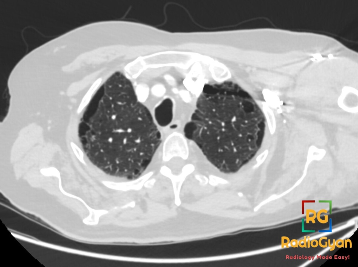
- Paraseptal (distal acinar) emphysema – alveolar destruction at the periphery of the acinus, abutting the pleura and interlobular septa.
- Single subpleural row of thin-walled lucencies along the pleura, fissures and septa, with upper lobe predominance.
- Lucencies are typically <1 cm; spaces ≥1 cm are bullae, which may coalesce into giant bullae.
- Smoking is the usual cause; predisposes to spontaneous pneumothorax.
- Chief mimic is honeycombing – one layer, upper lobes and no fibrosis versus stacked layers, lung bases and reticulation.
Imaging findings
- CT: round or oval lucencies in a single row along the subpleural lung, interlobar fissures and interlobular septa; walls are thin and well defined, corresponding to the septa.
- CT: upper lobe, often posterior, predominance; may be isolated or combined with centrilobular emphysema; subpleural bullae are frequent.
- Radiograph: typically normal unless bullae are large enough to appear as peripheral avascular lucencies.
Differential diagnosis
- Honeycombing (UIP) – stacked rows of cysts, basal and subpleural, with reticulation and traction bronchiectasis.
- Centrilobular emphysema – lucencies centred within the secondary lobule, sparing a subpleural rim, without perceptible walls.
- Lymphangioleiomyomatosis – diffuse round thin-walled cysts in all zones without a zonal gradient.
- Pulmonary Langerhans cell histiocytosis – bizarre-shaped cysts with nodules, sparing the costophrenic angles.
Classification
- Fleischner Society emphysema subtypes: centrilobular, panlobular, paraseptal and irregular (cicatricial); paraseptal corresponds to distal acinar emphysema.
What to do next
- Smoking cessation; spirometry and DLCO may be normal in isolated disease.
- Counsel on pneumothorax risk from bulla rupture; bullectomy with pleurodesis for recurrent pneumothorax or a giant bulla compressing lung.
- Review the lower lobes for fibrosis – emphysema with basal honeycombing suggests combined pulmonary fibrosis and emphysema (CPFE), in which DLCO is markedly reduced.
References
- Yuan Q, Yang X, Gu Y, Nie K, Liu J, Yue S, et al. Ultra-Low-Dose Photon-counting Detector CT for Emphysema Assessment: A Head-to-Head Comparative Study with Low-Dose CT. Radiology. 2026;318(1):e251609. PMID: 41557898.
- Cabanne E, Revel MP. Post-COVID-19 Vanishing Paraseptal Emphysema. Radiology. 2021;299(2):E249. PMID: 33620292.
- Sharma N, Justaniah AM, Kanne JP, Gurney JW, Mohammed TL. Vanishing lung syndrome (giant bullous emphysema): CT findings in 7 patients and a literature review. J Thorac Imaging. 2009;24(3):227-30. PMID: 19704328.

Improve Content And Help Your Colleagues!
Found an error in the post? Please let us know using our contact page and we will update it with due credits!
If you wish to contribute radiological images for this case (or any other article on the website), you can submit them here and you will be duly credited: Submit radiology case