


Key points
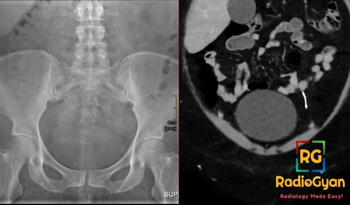
- A correctly placed Essure microinsert appears on CT as a curvilinear metallic density spanning the uterotubal junction at the uterine cornua; the normal fallopian tube course is not delineated on CT, so the device may appear to extend beyond the uterine contour.
- A short curvilinear metallic foreign body in the peritoneal cavity means the insert has extruded, from tubal perforation at the time of placement or distal migration through the tube – a rare complication, reported in about 0.04% of placements.
- Overly distal placement increases the risk of tubal perforation and peritoneal extrusion, and left-sided inserts are more often reported to migrate than right-sided ones.
- Extruded devices cause chronic pelvic pain and can induce peritoneal adhesions with potential small bowel obstruction, so they require surgical removal; CT is the adjunct used to locate the device or fragments before laparoscopic retrieval.
- HSG is the test of choice for confirming device position and tubal occlusion; on ultrasound the insert is an echogenic linear structure with myometrium surrounding it, and on MRI it produces susceptibility artifact at the cornua.
- Essure sales stopped in 2017 and US use ended in 2018, but many women still carry the inserts – assess device position whenever it is seen incidentally on CT, ultrasound, MRI or PET-CT.

Improve Content And Help Your Colleagues!
Found an error in the post? Please let us know using our contact page and we will update it with due credits!
If you wish to contribute radiological images for this case (or any other article on the website), you can submit them here and you will be duly credited: Submit radiology case