
Hypoxic- Ischemic Encephalopathy (HIE)
Pathophysiology of HIE
- Causes in adults:
- Drowning
- Asphyxiation
- Cardiac arrest
- Cerebrovascular accident
- Selectively vulnerable areas of the brain: For detailed pathophysiology cascade click here.
- Areas with highest concentrations of glutamate or other excitatory amino acid receptors
- Areas of the brain with the greatest energy demands.
- Mild to moderate ischemic insult: Watershed territory infarcts.
- Severe ischemic insult:Basal ganglia and thalami.
- Cerebral cortex
- Cerebellum
- Hippocampi
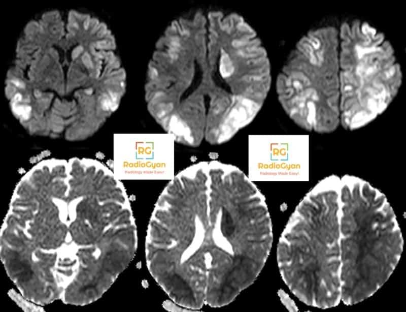
Imaging features of Hypoxic ischemic encephalopathy
- Changes are seen earliest on DWI (few hours) as areas of restricted diffusion.
- T2W images positive in the early subacute period (24 hours–2 weeks),
- DWI can pseudonormalize by the end of the 1st week while T2W abnormalities persist.
- The outcome is relatively poor.
| Imaging findings associated with relatively better outcome: | Imaging findings associated with relatively poor outcome: |
|
|
- Imaging algorithm:Click here
DICOM scrollable case
References and further reading:
- RSNA: Hypoxic-Ischemic Brain Injury Imaging Findings from Birth to Adulthood
- Muttikkal, TJ, Wintermark, M. MRI patterns of global hypoxic-ischemic injury in adults. J Neuroradiol 2013; 40: 164–171.

Improve Content And Help Your Colleagues!
Found an error in the post? Please let us know using our contact page and we will update it with due credits!
If you wish to contribute radiological images for this case (or any other article on the website), you can submit them here and you will be duly credited: Submit radiology case
Wow