


Key points
- Normal course – a femoral PICC should ascend the external and common iliac veins with its tip in the IVC or at the cavoatrial junction
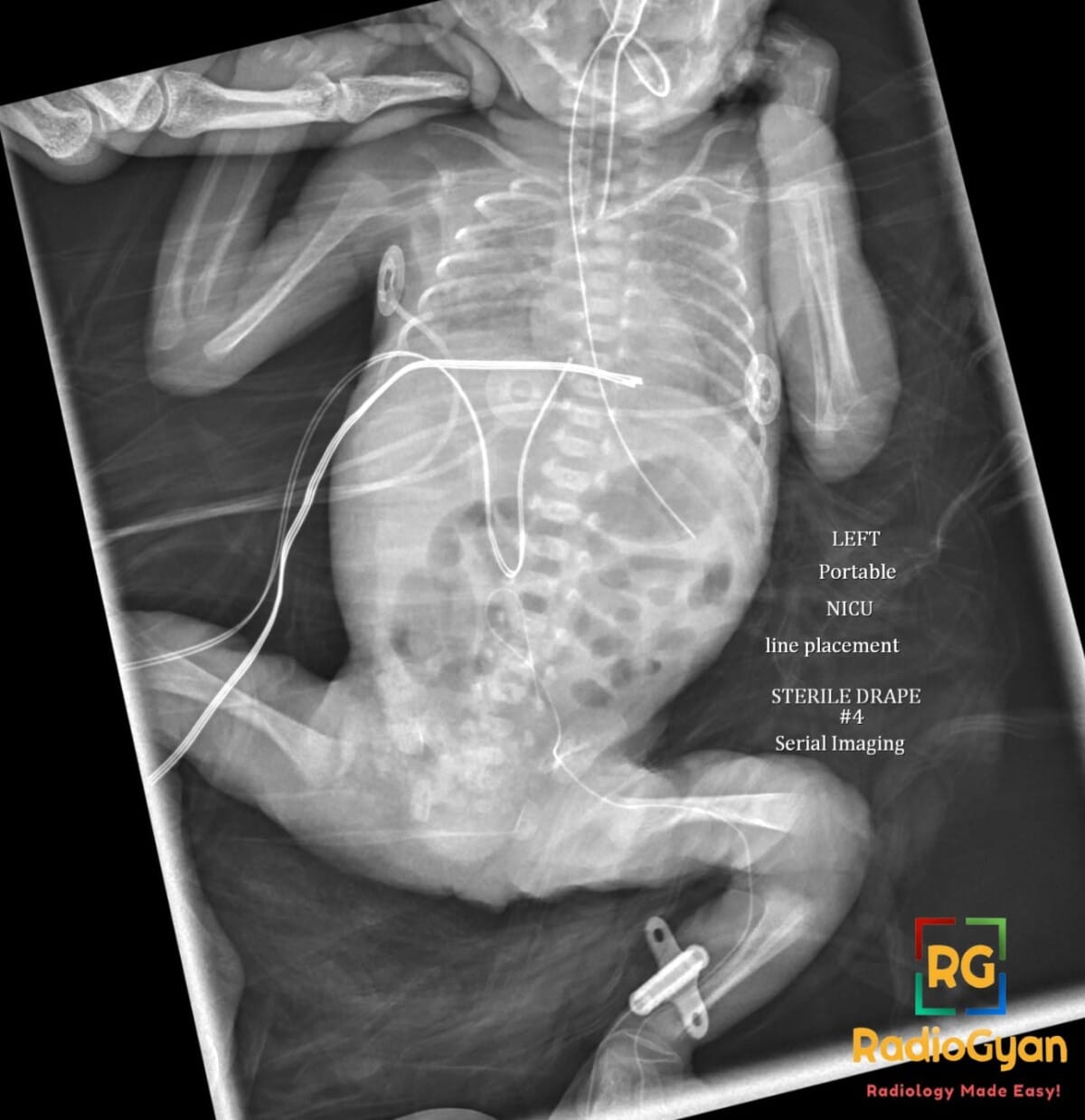
- Radiograph – catheter doubles back into a hairpin coil over the pelvis at the level of the iliac veins, the tip never reaching the IVC
- Trace to the tip – follow the coil to its end so it is not mistaken for entry into the contralateral iliac vein or an ascending lumbar vein
- Left-sided access – the catheter must cross the left common iliac vein beneath the right common iliac artery (May-Thurner point), a classic site of impingement and buckling
- Complications – catheter dysfunction, catheter-related thrombosis, phlebitis from sclerosant infusing small pelvic veins, vessel wall erosion or perforation
- Management – do not use the line until position is confirmed; reposition under fluoroscopy with guidewire manipulation, or withdraw and re-site

Improve Content And Help Your Colleagues!
Found an error in the post? Please let us know using our contact page and we will update it with due credits!
If you wish to contribute radiological images for this case (or any other article on the website), you can submit them here and you will be duly credited: Submit radiology case