


32-year-old woman undergoing evaluation for infertility. What is your diagnosis?
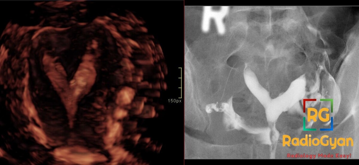
Diagnosis and teaching points:
Diagnosis: Septate uterus. 3D ultrasound and HSG demonstrate a septum dividing the uterine cavity.
Key facts for board exams:
- A normal convex fundal contour with an internal midline septum indicates septate uterus, whereas a deep external fundal cleft over 1 cm indicates bicornuate uterus.
- HSG alone is about 55% accurate for septate versus bicornuate uterus, so 3D ultrasound must confirm any positive HSG before surgery.
- ASRM 2021 criteria call for a septum longer than 1 cm, measured from the interostial line, with a leading-edge angle under 90 degrees.
- Randomized trial data show septum resection gives no live birth benefit over expectant management.
Detailed teaching points:
- Clinical: Most common Müllerian anomaly, usually found during infertility workup, recurrent pregnancy loss, or an abnormal pregnancy course. Breech presentation, preterm birth, and growth restriction are associated. Renal evaluation is not routinely required for an isolated septum.
- Etiology/Pathophys: Müllerian ducts fuse normally, but the central uterovaginal septum fails to resorb. The septum is poorly vascularized fibrous and muscular tissue, which impairs placentation and raises first-trimester loss risk.
- Radiograph: HSG shows a thin midline filling defect separating two symmetric cavities. Both horns usually opacify, and the fundal contour cannot be assessed.
- US: The coronal plane of 3D transvaginal ultrasound shows a convex or minimally indented external fundus with a deep internal indentation. 3D ultrasound has about 98% sensitivity and 100% specificity. The CUME threshold is indentation depth of 10 mm or more with an angle under 140 degrees.
- MRI: T2 shows a convex external fundal contour with an internal indentation over 1.5 cm. A thin septum is markedly hypointense (fibrous), while a thick septum is isointense to myometrium (muscular). Oblique planes along the long axis are required, since standard axial imaging can miss the fundal contour.
- Signs: V-shaped cavity sign – two diverging cavities with intercornual angle under 75 degrees on HSG; normal convex fundal contour sign – convex external fundus with a deep internal septum on coronal 3D ultrasound or oblique MRI.
- Frameworks: AFS 1988 classification places septate uterus in Class V. ESHRE/ESGE 2013 classifies it as U2 and overdiagnoses it by absorbing arcuate uteri. ASRM MAC 2021 and CUME criteria are more conservative.
- DDx: Bicornuate uterus – deep external fundal cleft, intercornual angle over 105 degrees, and HSG cannot reliably separate it from septate; uterus didelphys – two separate uterine bodies with two serosal surfaces, often confused with a complete septate uterus with duplicated cervix; arcuate uterus – normal contour with internal indentation under 1 cm and obtuse angle over 90 degrees.
- Tx: Expectant management for asymptomatic women and adolescents. Hysteroscopic metroplasty for recurrent pregnancy loss in shared decision-making, selected pre-IVF cases, and symptomatic complete septum with cervical or vaginal septum. Laparoscopy may confirm the fundal contour. Follow-up sonohysterography or 3D ultrasound assesses resection completeness and adhesions.
OSCE Questions
Question: Which modality is the best noninvasive test for this condition?
3D transvaginal ultrasound, with sensitivity about 98% and specificity about 100%.
Question: Which external fundal contour finding distinguishes this condition from a bicornuate uterus?
A convex, flat, or minimally indented (less than 1 cm) fundal contour.
Question: What intercornual angle on HSG suggests this condition?
An angle less than 75 degrees, forming a V or Y shape.
Question: Which Müllerian classification system overdiagnoses this condition by counting arcuate uteri as septate?
The ESHRE/ESGE classification (U2 class), which uses a threshold of internal indentation greater than 50% of uterine wall thickness.
Question: What is the most common uterine anomaly associated with recurrent pregnancy loss?
Septate uterus, with a reported miscarriage relative risk of 2.9 versus controls.
MCQ Questions
1. On 3D transvaginal ultrasound coronal imaging, which fundal contour pattern defines a septate uterus rather than a bicornuate uterus?
A. Deep external fundal cleft greater than 1 cm
B. Flat external fundus with absent internal indentation
C. Convex external fundus with deep internal indentation
D. Concave external fundus with shallow internal indentation
Answer: C. Convex external fundus with deep internal indentation. A convex or flat external fundus with a deep internal indentation reflects completed lateral fusion with failed septal resorption, whereas a deep external cleft indicates bicornuate morphology.
2. Using ASRM 2021 3D ultrasound criteria, which septum measurement defines a septate uterus?
A. Indentation depth below 1 cm with angle above 90 degrees
B. Septum length above 1 cm with angle below 90 degrees
C. Septum length below 1 cm with angle above 90 degrees
D. Indentation depth above 1 cm with angle above 90 degrees
Answer: B. Septum length above 1 cm with angle below 90 degrees. ASRM 2021 defines septate uterus by septum length above 1 cm with leading-edge angle below 90 degrees, while an obtuse angle with shallow depth indicates arcuate or normal.
3. Which best-performing noninvasive modality shows the external fundal contour needed to separate a septate uterus from a bicornuate uterus?
A. Hysterosalpingography
B. Standard axial pelvic MRI
C. Two-dimensional transvaginal ultrasound
D. Three-dimensional transvaginal ultrasound coronal reconstruction
Answer: D. Three-dimensional transvaginal ultrasound coronal reconstruction. Coronal three-dimensional transvaginal ultrasound displays serosal and endometrial contours together, which HSG cannot show and standard axial MRI often misses.
4. On ultrasound of a septate uterus, where does a gestational sac sit when implanted on the septum?
A. Centered on the midline septal band
B. Eccentric within the right endometrial cavity
C. Within the endocervical canal
D. Embedded in the right fallopian tube
Answer: A. Centered on the midline septal band. Implantation on the septum places the gestational sac centered on the midline band, a position associated with defective placentation and frequent first-trimester loss.
5. Which ASRM subcategory describes an asymmetric septate cavity that forms a blind cavity?
A. Partial septate uterus
B. Robert’s uterus
C. Normal or arcuate uterus
D. Complete septate uterus with duplicated cervices
Answer: B. Robert’s uterus. Robert’s uterus is the ASRM subcategory with an asymmetric septum producing a blind cavity, distinct from symmetric partial and complete septate forms.
Check out more such cases:






Improve Content And Help Your Colleagues!
Found an error in the post? Please let us know using our contact page and we will update it with due credits!
If you wish to contribute radiological images for this case (or any other article on the website), you can submit them here and you will be duly credited: Submit radiology case