


Older adult with low back pain radiating down the right leg. What is your diagnosis?
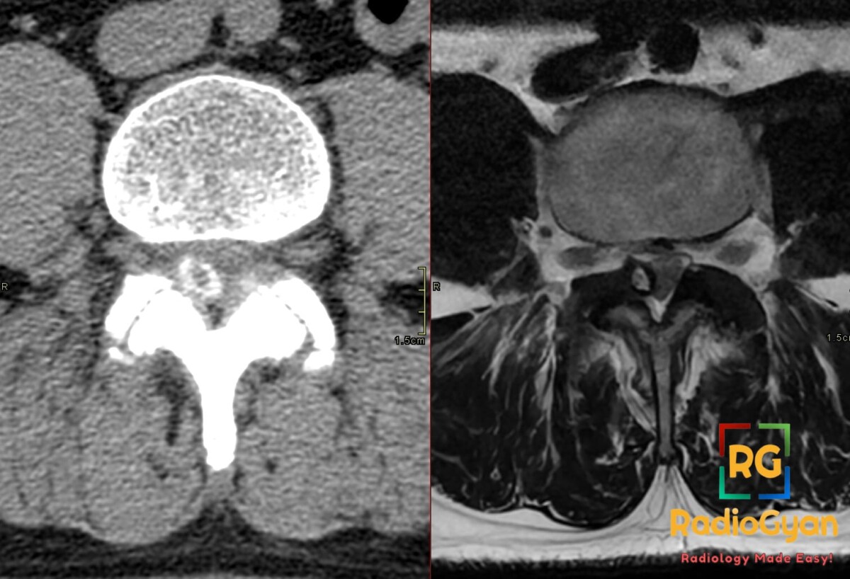
Diagnosis and teaching points:
Diagnosis: Peripherally calcified synovial cyst in the right half of the lumbar spinal canal. Mass effect on the right subarticular zone and the traversing right L5 nerve root.
Key facts for board exams:
- This is an extradural cyst arising from a degenerated facet joint capsule. The most common level is L4-L5, followed by L5-S1.
- Anteromedial extension into the lateral recess compresses the traversing root, which is L5 at L4-L5. Foraminal extension affects the exiting root instead.
- A T2 hypointense rim is characteristic. It may reflect calcification, hemorrhage, or the synovial lining itself.
- A completely calcified cyst can be dark on both T1 and T2 and hide its cystic nature. CT confirms the mineralization and shows the facet origin.
- These cysts are strongly linked to degenerative spondylolisthesis and segmental instability, so look for listhesis at the same level.
Detailed teaching points:
- Clinical: Usually seen in the sixth or seventh decade, with no consistent sex predilection. Patients present with back pain, unilateral radiculopathy, or neurogenic claudication; cauda equina syndrome is rare. Intracystic hemorrhage causes abrupt worsening. Nearly half of cysts found on lumbar MRI are incidental.
- Etiology/Pathophys: Facet arthropathy and segmental hypermobility weaken the joint capsule. Synovium then herniates as an outpouching that communicates with the joint. The wall can later fibrose, bleed, calcify, or rarely ossify.
- CT: Shows a well-defined posterolateral extradural mass abutting an arthritic facet. Rim calcification is more common than complete calcification. Intracystic gas or a facet vacuum phenomenon may be present. Rare pressure erosion of the lamina can mimic tumor. CT arthrography shows contrast filling the cyst from the joint.
- MRI: MRI is the preferred study, and axial images best show lateral recess compression. The cyst is typically T1 low and T2 bright. Protein or blood raises T1 signal and lowers T2 signal. After gadolinium there is thin peripheral enhancement and no solid component.
- Signs: Aunt Minnie appearance – a smooth posterolateral extradural cyst contiguous with an arthritic facet, indenting the thecal sac and lateral recess.
- Frameworks: NSURG grading combines canal compromise on axial T2 with listhesis. Grades I-III have less than 15% listhesis (grade I occupies less than 25% of the canal; grade II, less than 50%). Grade IV is 15% or more listhesis with less than 50% compromise. Grade V is 15% or more listhesis with more than 50% compromise.
- DDx: Sequestered disc fragment – connects to the disc, has no facet relationship, and lies anterior to the ligamentum flavum; ganglion cyst – identical on imaging and lacks a synovial lining only on histology; calcified ligamentum flavum – CT shows ligament origin rather than facet origin; cystic nerve sheath tumor – centered on the root, with an enhancing solid component.
- Tx: Incidental or mild cases are managed conservatively, and spontaneous regression can occur. Options for symptomatic cysts are CT-guided facet steroid injection, aspiration, or rupture, though calcified cysts are hard to puncture. Refractory pain or a progressive deficit warrants decompression with cyst excision, with fusion added if instability is present.
OSCE Questions
Question: What modality is preferred for detection of this condition?
MRI, showing facet-adjacent extradural mass and root compression.
Question: What is characteristic peripheral T2 finding in this condition?
Peripheral low-signal rim from synovial lining, calcification or hemorrhage.
Question: Which modality best confirms suspected calcification in this condition?
CT, showing rim or complete high-attenuation mineralization adjacent to degenerated facet.
Question: What grading system incorporates canal compromise and listhesis for this condition?
NeuroSpine Surgery Research Group (NSURG) grading system.
Question: Which traversing root is compressed by medial L4-L5 extension?
Traversing L5 nerve root in the subarticular lateral recess.
MCQ Questions
1. In known lumbar facet synovial cyst, which MRI feature most supports capsule origin?
A. Diffuse homogeneous enhancement
B. Peripheral low signal rim
C. Central fluid fluid level
D. Anterior disc continuity
Answer: B. Peripheral low signal rim. A T2 low signal peripheral rim reflects synovial lining, calcification or hemorrhage and favors facet cyst.
2. In known lumbar synovial cyst, which feature best distinguishes it from sequestered disc fragment?
A. Facet joint adjacency
B. Anterior epidural location
C. Disc space narrowing
D. Vertebral edema pattern
Answer: A. Facet joint adjacency. Facet adjacency or communication with cystic rim favors synovial cyst over disc related anterior fragment.
3. According to NSURG grading of lumbar synovial cyst, Grade V requires which combination?
A. Under 25 percent canal without listhesis
B. Under 50 percent canal without listhesis
C. 15 percent listhesis plus under 50 percent canal
D. 15 percent listhesis plus over 50 percent canal
Answer: D. 15 percent listhesis plus over 50 percent canal. NSURG Grade V is at least 15 percent listhesis with greater than 50 percent canal compromise.
4. In suspected calcified lumbar synovial cyst, which modality best confirms mineralization?
A. Computed tomography
B. Magnetic resonance imaging
C. Diagnostic ultrasound
D. Bone scintigraphy
Answer: A. Computed tomography. CT depicts rim or complete calcification, gas vacuum and osseous remodeling adjacent to degenerated facet.
5. In known lumbar synovial cyst with acute deterioration, which imaging finding indicates hemorrhagic complication?
A. T2 uniform hyperintensity only
B. Absent peripheral enhancement
C. T1 hyperintensity within cyst
D. Adjacent facet vacuum alone
Answer: C. T1 hyperintensity within cyst. Proteinaceous material or hemorrhage can make a cyst T1 isointense or hyperintense instead of T1 low.
Check out more such cases:

Improve Content And Help Your Colleagues!
Found an error in the post? Please let us know using our contact page and we will update it with due credits!
If you wish to contribute radiological images for this case (or any other article on the website), you can submit them here and you will be duly credited: Submit radiology case