


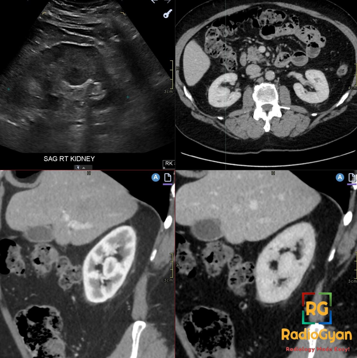
Incidental finding in a 55-year-old male. What is your diagnosis?
Diagnosis and teaching points:
Diagnosis: Hypertrophied column of Bertin in the right kidney interpolar region, suspected as a mass on ultrasound and confirmed on subsequent multiphase CT.
Key facts for board exams:
- Benign pseudotumor of normal cortex projecting into the renal sinus; not a neoplasm (described by Bertin, 1744).
- Most often interpolar (mid-kidney), between upper and middle calyces; left-sided predominance, may be bilateral.
- Central discriminator: the lesion is continuous with cortex and behaves like normal parenchyma on every modality.
- US shows a preserved outer renal contour with Doppler flow matching adjacent parenchyma, arguing against a true mass.
- Classic pitfall: unnecessary biopsy or nephrectomy for this benign anatomic variant.
Detailed teaching points:
- Clinical: Incidental and asymptomatic at any age, including children; no established sex predilection, risk factor, or syndromic association.
- Etiology/Pathophys: Normal cortex extending between pyramids into the sinus, likely persistent junctional parenchyma from incomplete fusion of embryonic sub-kidneys; histologically normal tissue containing interlobular vessels.
- US: Well-defined projection, isoechoic to mildly hyperechoic, sometimes engulfing a papilla; CEUS enhancement parallels surrounding cortex.
- CT: Isoattenuating to cortex on noncontrast, with homogeneous enhancement matching cortex on corticomedullary and nephrographic phases; collecting system may be distorted; no necrosis, calcification, or capsule.
- MRI: T1 and T2 signal isointense to cortex; dynamic post-contrast enhancement and washout parallel adjacent parenchyma; useful when iodinated contrast is unsuitable.
- Signs: Split sinus sign – cortical projection indents or divides central sinus echoes; claw-like sinus – sinus echoes partly surround the column; truncated calyx with opposing renal-margin dimple on excretory urography.
- DDx: Renal cell carcinoma (classic trap) – independent mass with distinct interface and enhancement differing from cortex; persistent fetal lobulation – external contour indentations, not internal projection; dromedary hump – focal lateral bulge from splenic impression; focal compensatory hypertrophy – functioning tissue lacking junctional sinus anatomy.
- Tx: No disease-directed treatment; reassure and document. If atypical, characterize with CEUS, multiphase CT, or MRI (radiation-free options preferred in children) before invasive steps; follow-up individualized until stability is proven.
OSCE Questions
Question: Which modality best characterizes this pseudotumor when ultrasound is indeterminate?
Multiphase contrast-enhanced CT — confirms cortical continuity and enhancement matching normal renal cortex.
Question: Which named ultrasound sign supports this diagnosis?
Split sinus sign — the cortical projection indents or divides the central renal sinus echoes.
Question: Which adult malignancy is the principal differential diagnosis?
Renal cell carcinoma (RCC).
Question: What single feature most reliably distinguishes this condition from a true mass?
Continuity with renal cortex and enhancement identical to normal parenchyma.
Question: What treatment is indicated after confident diagnosis?
None — reassurance and documentation; avoid biopsy or surgery.
MCQ Questions
1. Which ultrasound sign describes the cortical projection dividing or splaying the central renal sinus echoes in hypertrophied column of Bertin?
A. Opposing renal-margin dimple
B. Split sinus sign
C. Truncated calyx sign
D. Dromedary hump
Answer: B. Split sinus sign. The split sinus sign describes normal cortical tissue projecting inward and dividing or splaying the central renal sinus echoes; it is supportive but not independently pathognomonic.
2. Which multiphase CT feature best distinguishes a hypertrophied column of Bertin from renal cell carcinoma?
A. Heterogeneous enhancement with central necrosis
B. Distortion of the outer renal contour
C. Hypoenhancement relative to cortex on nephrographic phase
D. Homogeneous enhancement matching cortex with preserved renal contour
Answer: D. Homogeneous enhancement matching cortex with preserved renal contour. The column is continuous with cortex and enhances identically across phases while preserving the outer contour, whereas renal cell carcinoma typically forms a distinct mass with differing enhancement.
3. In a child with an indeterminate renal pseudomass on ultrasound, which radiation-free modality confirms enhancement paralleling the renal cortex?
A. Contrast-enhanced ultrasound
B. 99mTc-DMSA scintigraphy
C. Multiphase contrast CT
D. Excretory urography
Answer: A. Contrast-enhanced ultrasound. CEUS demonstrates enhancement and washout paralleling renal cortex without ionizing radiation, making it a preferred problem-solving tool in children, unlike DMSA scintigraphy, CT, and excretory urography.
4. On nephrographic phase CT, a hypertrophied column of Bertin demonstrates which enhancement pattern?
A. Rim enhancement with central low attenuation
B. Progressive centripetal enhancement with delayed washout
C. Isoattenuation to adjacent renal cortex
D. Marked hypoenhancement relative to parenchyma
Answer: C. Isoattenuation to adjacent renal cortex. The column consists of normal functioning parenchyma, so it remains isoattenuating to cortex on nephrographic phase without necrosis, rim enhancement, or independent mass behavior.
5. Which feature distinguishes a hypertrophied column of Bertin from persistent fetal lobulation?
A. Smooth external indentations between renal lobes
B. Inward cortical projection into the renal sinus
C. Underlying pyramid within each surface lobulation
D. Focal bulge of the lateral renal border
Answer: B. Inward cortical projection into the renal sinus. Hypertrophied column of Bertin projects internally toward the sinus with a preserved external contour, whereas persistent fetal lobulation causes smooth external indentations with underlying pyramids.
Check out more such cases:

Improve Content And Help Your Colleagues!
Found an error in the post? Please let us know using our contact page and we will update it with due credits!
If you wish to contribute radiological images for this case (or any other article on the website), you can submit them here and you will be duly credited: Submit radiology case