


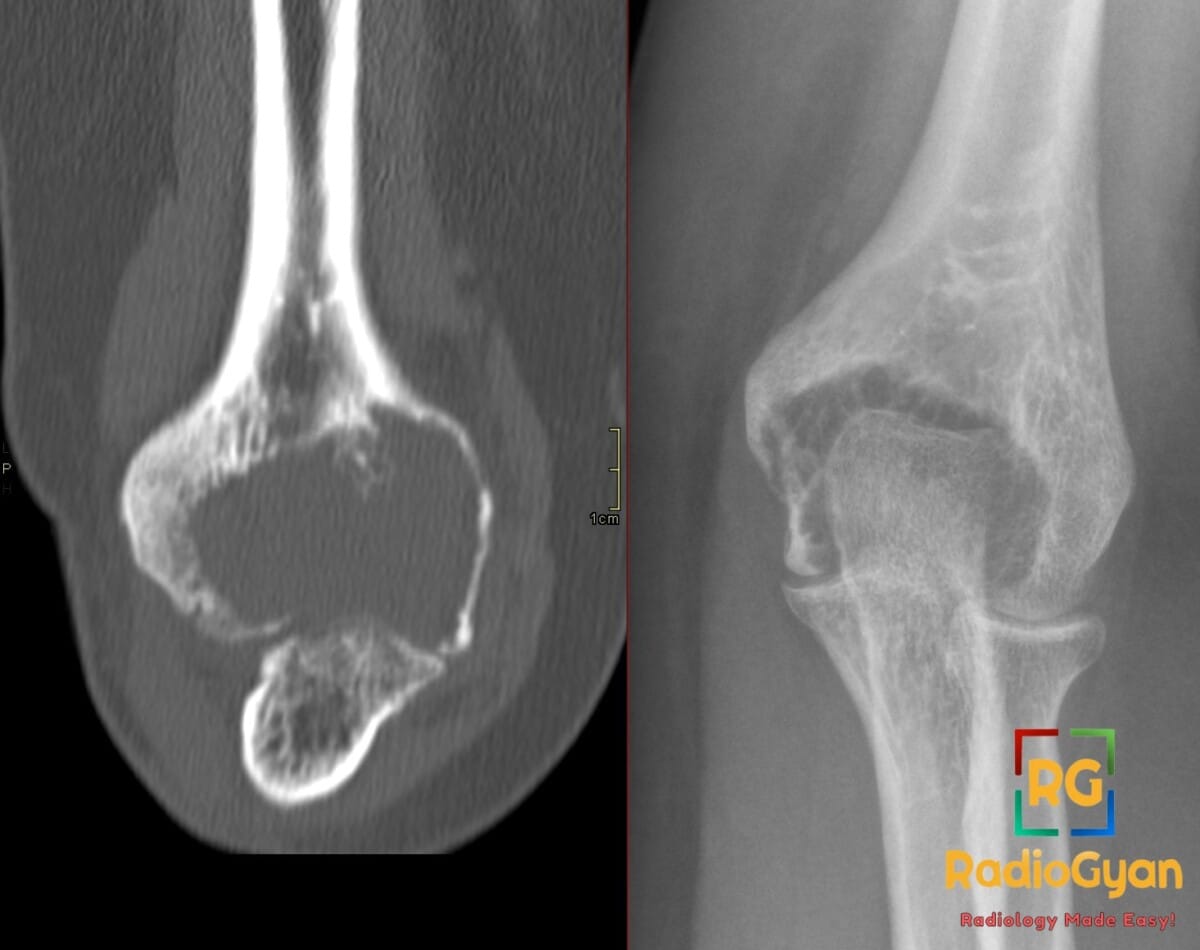
30-year-old female with progressive elbow pain. What is your diagnosis?
Diagnosis and teaching points:
Diagnosis: Distal humeral giant cell tumor in a skeletally mature patient.
Key facts for board exams:
- Arises almost exclusively in skeletally mature patients aged 20–40 years, extending from the metaphysis directly to the subchondral articular surface.
- Driven by a somatic H3F3A G34W mutation in neoplastic mononuclear stromal cells that recruit non-neoplastic osteoclasts via RANKL.
- Classically presents as an eccentric, geographic osteolytic lesion lacking a sclerotic margin or internal mineralized matrix.
- Frequently displays low T1 and T2 signal foci on MRI representing hemosiderin deposition from recurrent intratumoral hemorrhage.
- Classified by the WHO as intermediate malignant due to local aggressiveness and rare benign pulmonary metastasis.
Detailed teaching points:
- Clinical: Slight female predilection; presents with progressive joint pain, limited elbow range of motion, swelling, or pathological fracture; associated with Paget disease of bone.
- Etiology/Pathophys: Neoplastic stromal cells stimulate osteoclastogenesis via RANK-RANKL pathway signaling, driving enzymatic bone resorption via cathepsin K and matrix metalloproteinases into an expansile osteolytic lesion.
- Radiograph: Demonstrates endosteal scalloping, cortical expansion, and cortical thinning abutting the subchondral plate without periosteal reaction unless a pathological fracture is present.
- CT: Delineates exact cortical breach, subchondral bone plate thinning, and residual trabeculae to assist operative planning and joint salvage.
- MRI: Demonstrates heterogeneous hyperintensity on T2/STIR, avid enhancement of solid vascular components, and assesses soft-tissue extension, marrow involvement, and joint capsule invasion.
- Nuc Med: Intense FDG avidity on PET/CT (mean SUVmax around 9) mimicking high-grade malignancy; bone scintigraphy demonstrates increased peripheral radiotracer uptake.
- Signs: Soap-bubble appearance – expansile osteolysis with residual bony pseudoseptations; Paintbrush borders sign – brush-like infiltrating margins on T1 MRI indicating higher recurrence risk.
- Frameworks: Campanacci grading stages lesions from 1 (intact cortex), to 2 (expanded/thinned cortex), to 3 (cortical destruction with soft-tissue extension); Enneking staging categorizes benign-aggressive musculoskeletal tumors.
- DDx: Aneurysmal bone cyst – predominantly cystic lacking solid enhancing tissue; Chondroblastoma – skeletally immature with chondroid calcifications; Brown tumor – associated with elevated PTH and hypercalcemia; Telangiectatic osteosarcoma – thick nodular septa with malignant osteoid.
- Tx: Intralesional curettage with PMMA cementation and local adjuvants; denosumab for unresectable or recurrent disease; wide en bloc resection if the articular surface cannot be preserved.
OSCE Questions
Question: Which driver gene mutation is characteristic of this pathology?
H3F3A mutation, most commonly encoding the p.Gly34Trp (G34W) substitution.
Question: What radiographic grading system is traditionally used for this pathology?
Campanacci radiographic grading system.
Question: What MRI sign on T1-weighted imaging indicates increased risk of local recurrence?
The paintbrush borders sign.
Question: Which targeted monoclonal antibody is used for medical management of this tumor?
Denosumab, an inhibitor of receptor activator of nuclear factor kappa-B ligand (RANKL).
Question: What is the most common site of distant metastasis in this condition?
The lungs (pulmonary metastasis).
MCQ Questions
1. Which specific driver mutation is most frequently identified in the neoplastic mononuclear stromal cells of giant cell tumor of bone?
A. IDH1 R132H
B. H3F3A G34W
C. GNAS R201H
D. USP6 rearrangement
Answer: B. H3F3A G34W. Approximately 90–95% of giant cell tumors of bone harbor an H3F3A driver mutation, most commonly the p.Gly34Trp (G34W) substitution in neoplastic mononuclear stromal cells.
2. In the Campanacci radiographic grading system for giant cell tumor of bone, which finding defines a Grade 3 lesion?
A. Sclerotic margin with intact cortex
B. Secondary fluid-fluid level formation
C. Extensive cortical breakthrough and soft-tissue extension
D. Cortical thinning without contour deformation
Answer: C. Extensive cortical breakthrough and soft-tissue extension. Campanacci Grade 3 lesions exhibit fuzzy margins, marked cortical destruction, and soft-tissue extension beyond the bone without a constrained cortical rim.
3. On MRI of giant cell tumor of bone, which margin characteristic is associated with a significantly increased risk of post-curettage local recurrence?
A. Paintbrush borders sign
B. Fallen fragment sign
C. Double line sign
D. Target sign
Answer: A. Paintbrush borders sign. The paintbrush borders sign on MRI represents permeative micro-trabecular osteolytic infiltration beyond the main tumor boundary and correlates with higher local recurrence after curettage.
4. When differentiating a giant cell tumor from a primary aneurysmal bone cyst on contrast-enhanced MRI, which feature favors giant cell tumor?
A. Exclusively thin enhancing septa
B. Complete absence of internal enhancement
C. Peripheral non-enhancing cystic spaces
D. Prominent avidly enhancing solid components
Answer: D. Prominent avidly enhancing solid components. Giant cell tumors demonstrate prominent, avidly enhancing solid stromal components, whereas primary aneurysmal bone cysts consist predominantly of fluid-filled spaces with only thin enhancing internal septations.
5. In a patient with giant cell tumor of bone receiving denosumab therapy, which imaging finding typically indicates treatment response?
A. Progressive permeative cortical osteolysis
B. Intralesional osteosclerosis and neocortex formation
C. Rapidly expanding non-calcified soft-tissue mass
D. Development of aggressive periosteal reaction
Answer: B. Intralesional osteosclerosis and neocortex formation. Anti-RANKL therapy with denosumab inhibits osteoclast-mediated osteolysis, promoting prominent intralesional mineralization, osteosclerosis, and reconstitution of a peripheral neocortical rim.
Check out more such cases:


Improve Content And Help Your Colleagues!
Found an error in the post? Please let us know using our contact page and we will update it with due credits!
If you wish to contribute radiological images for this case (or any other article on the website), you can submit them here and you will be duly credited: Submit radiology case