


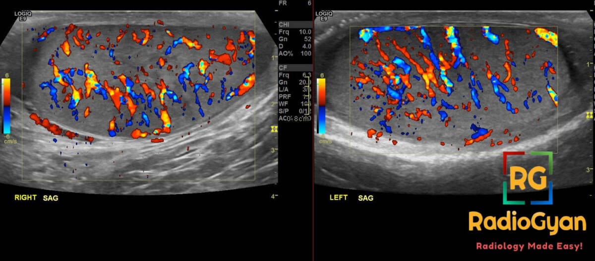
28-year-old male presenting with acute scrotal pain and swelling. What is your diagnosis?
Diagnosis and teaching points:
Diagnosis: Bilateral orchitis. Color Doppler ultrasound shows hyperemia in both testes.
Key facts for board exams:
- Bilateral testicular involvement most strongly suggests a systemic viral etiology, classically mumps, via hematogenous dissemination.
- Unlike testicular torsion, which shows reduced or absent perfusion, orchitis characteristically demonstrates testicular hyperemia on Doppler.
- Isolated orchitis is uncommon and typically hematogenous, whereas bacterial disease usually ascends from an adjacent epididymitis.
- Severe or viral inflammation can cause germ-cell destruction, subfertility, and irreversible testicular atrophy.
- Follow-up imaging is essential to ensure resolution and avoid misdiagnosing a necrotic testicular tumor as resolving infection.
Detailed teaching points:
- Clinical: Postpubertal males or young adults (19–35 years); presents with acute scrotal pain, swelling, fever, and erythema; risk factors include unprotected intercourse, bladder outlet obstruction, urinary instrumentation, and lack of vaccination.
- Etiology/Pathophys: Infectious pathogens induce vascular dilation, increased capillary permeability, and interstitial edema; inflammatory cytokine release disrupts the blood-testis barrier, impairing Leydig-cell function and triggering germ-cell apoptosis.
- Radiograph: Emphysematous epididymo-orchitis manifests as discrete pockets of radiolucent gas projecting over the scrotal soft tissues.
- US: Testicular enlargement with diffuse hypoechogenicity or heterogeneous echotexture; associated features include reactive hydroceles, internal septations, and scrotal-wall thickening.
- CT: Evaluates complicated disease, demonstrating perineal fat stranding, scrotal-wall edema, and loculated gas or nonenhancing fluid collections in necrotizing fasciitis.
- MRI: Demonstrates intermediate-to-low T1 and high T2/STIR signal from inflammatory edema, with intense parenchymal enhancement on post-contrast sequences.
- Nuc Med: Technetium-99m pertechnetate scintigraphy demonstrates diffusely increased hemiscrotal perfusion and radiotracer uptake throughout dynamic and tissue phases.
- Signs: Sunglasses view – comparative side-by-side testicular hypoechogenicity and hypervascularity; inferno sign – marked diffuse parenchymal color Doppler flow from intense hyperemia.
- DDx: Testicular torsion – absent or diminished Doppler flow and high-riding transverse testis; testicular tumor – persistent solid vascular mass unresponsive to antibiotics; testicular abscess – central avascular fluid with thick peripheral hyperemic rim.
- Tx: Supportive therapy with scrotal elevation and NSAIDs for viral disease; targeted antibiotic therapy for bacterial pathogens; repeat ultrasound within 3–7 days for non-improving symptoms.
OSCE Questions
Question: What surgical emergency must be immediately excluded in this clinical scenario?
Testicular torsion.
Question: What viral pathogen is classically associated with bilateral involvement in postpubertal males?
Mumps virus (paramyxovirus).
Question: What color Doppler ultrasound sign describes marked diffuse hypervascularity in this condition?
The inferno sign.
Question: Which two sexually transmitted pathogens most commonly cause ascending infection in young adults?
Chlamydia trachomatis and Neisseria gonorrhoeae.
Question: What long-term parenchymal complication commonly follows severe viral involvement?
Testicular atrophy, which can lead to subfertility or infertility.
MCQ Questions
1. What is the primary route of infection leading to isolated viral orchitis?
A. Retrograde lymphatic transit
B. Hematogenous dissemination
C. Ascending canalicular spread
D. Direct transluminal migration
Answer: B. Hematogenous dissemination. Isolated viral orchitis typically occurs via hematogenous dissemination, whereas acute pyogenic bacterial orchitis usually results from ascending infection from the urinary tract and epididymis.
2. Which color Doppler ultrasound sign denotes marked diffuse hypervascularity of the testicular parenchyma in acute orchitis?
A. Whirlpool sign
B. Yin-yang sign
C. Inferno sign
D. Blue dot sign
Answer: C. Inferno sign. The “inferno sign” on color Doppler ultrasound describes intense, diffuse parenchymal hypervascularity caused by severe active testicular inflammation.
3. In the ultrasound staging system for acute epididymo-orchitis, which feature categorizes disease as Stage IIIB?
A. Presence of an uncomplicated reactive hydrocele
B. Clear anatomical separation of gonadal margins
C. Complete loss of parenchymal arterial vascularity
D. Abscess exceeding 0.5 cm in size
Answer: D. Abscess exceeding 0.5 cm in size. Stage IIIB epididymo-orchitis is defined by indistinguishable testis and epididymis with one or more abscesses greater than 0.5 cm, serving as a primary surgical indication.
4. Which scrotal scintigraphy pattern on technetium-99m pertechnetate indicates acute testicular parenchymal involvement in epididymo-orchitis?
A. Diffusely increased hemiscrotal tracer uptake
B. Focal lateral crescentic tracer activity alone
C. Central photopenia with a hyperemic rim
D. Globally decreased spermatic cord perfusion
Answer: A. Diffusely increased hemiscrotal tracer uptake. Epididymitis alone classically demonstrates a lateral crescent of activity, whereas progression to orchitis produces diffuse hyperemia throughout the entire hemiscrotal parenchyma.
5. In acute bacterial orchitis, a lack of clinical improvement within what timeframe warrants repeat ultrasound to evaluate for abscess or infarction?
A. 12 to 24 hours
B. 48 to 72 hours
C. 5 to 7 days
D. 10 to 14 days
Answer: B. 48 to 72 hours. Patients failing to demonstrate clinical improvement after 48 to 72 hours of appropriate antibiotic therapy require repeat imaging to rule out suppurative complications like abscess or infarction.
Check out more such cases:

Improve Content And Help Your Colleagues!
Found an error in the post? Please let us know using our contact page and we will update it with due credits!
If you wish to contribute radiological images for this case (or any other article on the website), you can submit them here and you will be duly credited: Submit radiology case