


Key points
- Artificial urinary sphincter (AUS) is the gold standard surgical treatment for male stress urinary incontinence, most often after prostatectomy, with three components: a cuff around the bulbar urethra, a pressure-regulating balloon (reservoir) and a control pump in the scrotum.
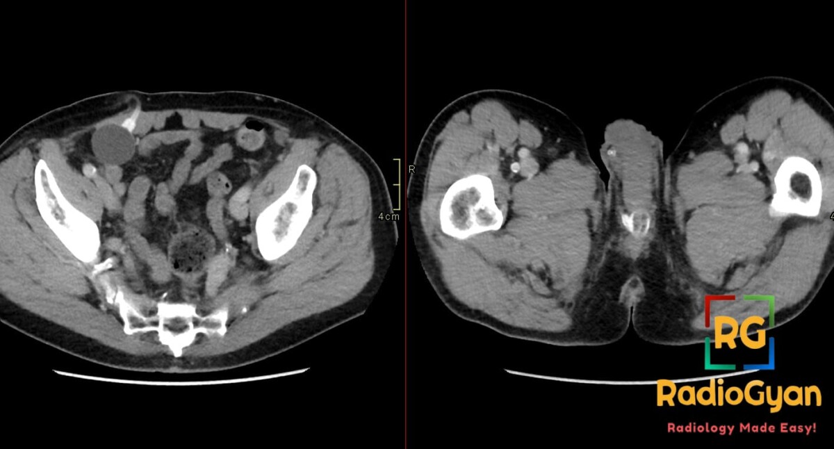
- CT: the cuff is a fluid-attenuation ring around the urethra at the penile base, the reservoir a rounded fluid-attenuation balloon in the prevesical space or high submuscular position that may project in either lower quadrant, and the pump an ovoid device in the scrotum, all joined by connecting tubing.
- Current reservoirs are filled with saline and appear fluid attenuation; older systems used radiopaque contrast, which allowed plain radiography to show the circuit and detect fluid loss.
- A baseline radiograph immediately after implantation is essential for later comparison; without one, reservoir volume loss is not detectable until about 50% has been lost.
- Complications: component malposition, mechanical failure with leak and loss of hydraulic fluid (the commonest cause of malfunction), infection, cuff erosion (contrast extravasates around the cuff on voiding cystourethrography) and urethral atrophy; CT or ultrasound can confirm fluid loss but not localize the leak.
- Gas around components is expected in the immediate postoperative period; gas with fat stranding beyond the early phase suggests infection, and CT is the modality of choice when a complication is suspected.
References
- Chorney ET, Ramchandani P, Jaffe WI, Siegelman ES. CT and MR Imaging Features of Artificial Urinary Sphincters, Penile Prostheses, and Other Devices in the Male Lower Genitourinary Tract. Radiographics. 2018;38(3):794-805. PMID: 29757723.
- Oda S, Kuno H, Hiyama T, Yamaguchi M, Sasaki T, Yajima S, et al. Radiologic feature of complications after artificial urinary sphincter implantation following total prostatectomy. Abdom Radiol (NY). 2024;49(7):2416-2427. PMID: 38849538.

Improve Content And Help Your Colleagues!
Found an error in the post? Please let us know using our contact page and we will update it with due credits!
If you wish to contribute radiological images for this case (or any other article on the website), you can submit them here and you will be duly credited: Submit radiology case