


Key points
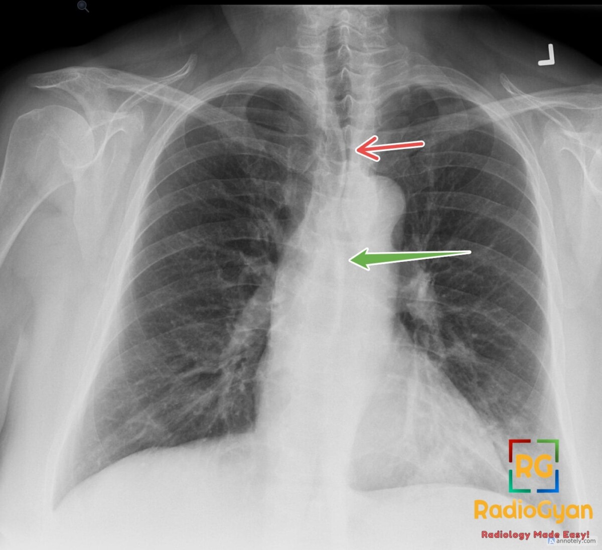
- Anterior junction line: four pleural layers (two visceral, two parietal) where the upper lobes appose anterior to the pericardium; visible on about 40% of upright frontal radiographs as an oblique line running downward from right to left at the level of the aortic arch.
- Posterior junction line: pleural apposition behind the trachea and esophagus, in front of the spine; seen more often than the anterior line, projected as a vertical line over the tracheal air column and extending above the clavicles down to the aortic arch.
- Both are normal potential spaces containing a little mediastinal fat; each is a hairline (≤1 mm) that lipomatosis may slightly widen in elderly or obese patients.
- Effacement or focal widening localizes adjacent disease: thymic lesions, thyroid mass, lymphoma, germ cell tumor for the anterior line; esophageal mass, lymphadenopathy, aortic disease, neurogenic tumor for the posterior line.
- Displacement matters as much as effacement – toward the lesion with volume loss (pulmonary hypoplasia, chronic scarring, atelectasis), away from it with anterior pneumothorax or congenital lobar overinflation (usually left upper lobe).
- The retrocardiac opacity is left lower lobe pneumonia, unrelated to the junctional lines; it hides behind the heart on the frontal view and classically produces the spine sign – loss of the normal progressive increase in vertebral lucency – on the lateral view.
References
- Yoon HK, Jung KJ, Han BK, Cho YA, Park JM, Lee KS, et al. Mediastinal interfaces and lines in children: radiographic-CT correlation. Pediatr Radiol. 2001;31(6):406-12. PMID: 11436887.
- Feragalli B, Mantini C, Patea RL, De Filippis F, Di Nicola E, Storto ML. Radiographic evaluation of mediastinal lines as a diagnostic approach to occult or subtle mediastinal abnormalities. Radiol Med. 2011;116(4):532-47. PMID: 21424313.

Improve Content And Help Your Colleagues!
Found an error in the post? Please let us know using our contact page and we will update it with due credits!
If you wish to contribute radiological images for this case (or any other article on the website), you can submit them here and you will be duly credited: Submit radiology case