
What causes Sister Mary Joseph sign in the umbilicus on clinical examination and imaging?
Let me know in the comments.
Click to reveal the answer
Answer:
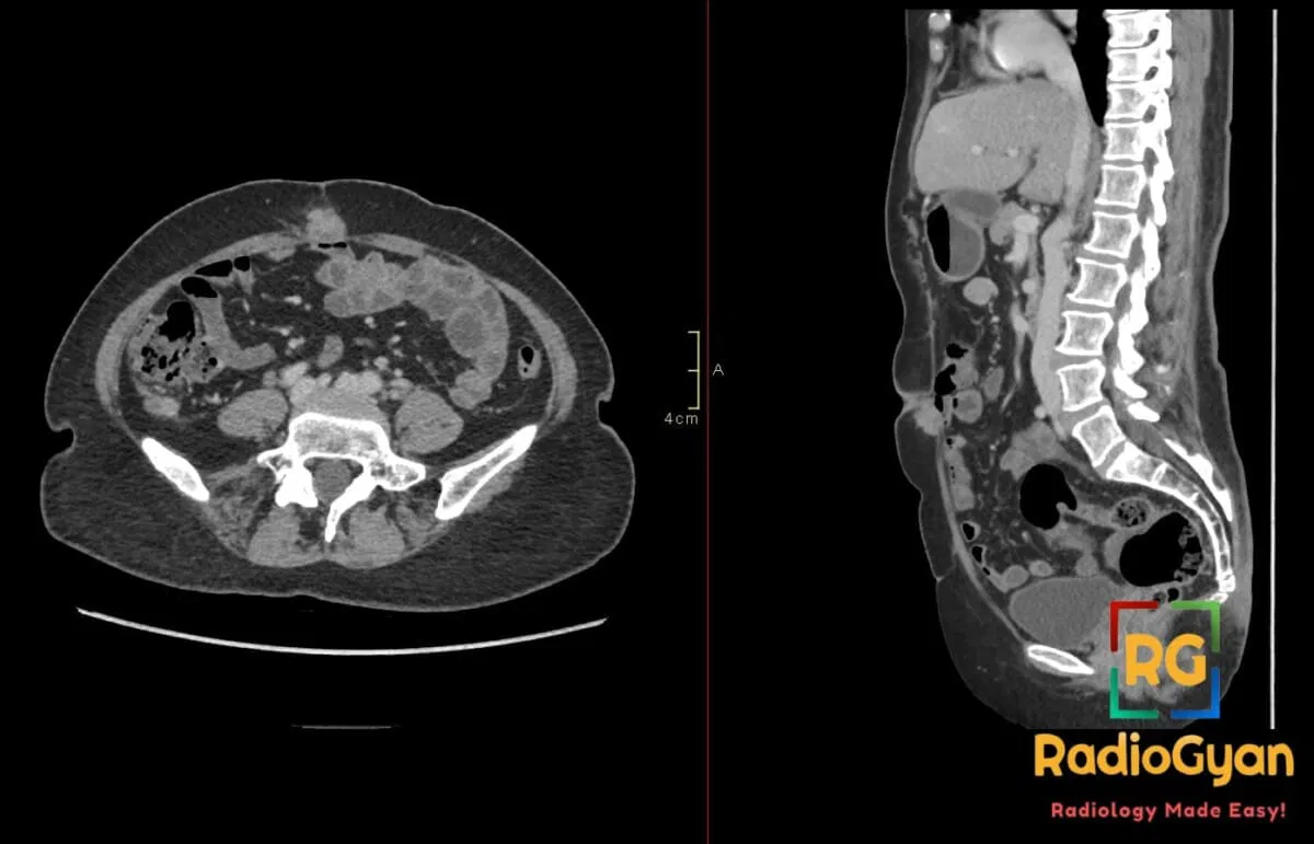
The Sister Mary Joseph sign is caused by metastatic malignant deposits in the umbilical region, originating from advanced intra-abdominal or pelvic malignancies, typically adenocarcinomas such as those arising from the stomach, pancreas, ovary, colon, or other abdominal organs.
It presents clinically as a palpable, often painful, firm periumbilical nodule or mass that may vary in size from a few millimeters up to several centimeters.
Imaging modalities like ultrasound or contrast-enhanced CT reveal a solid, enhancing umbilical lesion often associated with other signs of peritoneal carcinomatosis or omental metastasis. This sign indicates advanced metastatic disease and carries a poor prognosis.
Why is it called so?:
It is named after Sister Mary Joseph Dempsey, a surgical assistant to Dr. William J. Mayo at the Mayo Clinic in the early 20th century, who first noted the clinical association between a palpable umbilical nodule and advanced intra-abdominal malignancy. The eponym honors her observation of this metastatic umbilical lesion as a diagnostic clue.
Pathophysiology:
The sign develops through metastatic spread of malignant cells from a primary intra-abdominal or pelvic tumor to the umbilicus via lymphatic channels, venous pathways, or along the round ligament of the liver (a remnant of the fetal umbilical vein). Tumor cells invade the umbilical soft tissues, leading to the formation of a firm nodular mass. The umbilicus serves as a vulnerable anatomical site due to its rich vascular and lymphatic connections to the peritoneal cavity, allowing tumor implantation and growth at this superficial location.
Alternative names:
Sister Mary Joseph nodule, Sister Mary Joseph node
Other associated named signs:
No other named signs are directly associated with this lesion, but it often occurs in the context of signs of peritoneal carcinomatosis such as ascites or omental caking.
Access all radiology signs posted so far: https://radiogyan.com/radiology-signs/

Improve Content And Help Your Colleagues!
Found an error in the post? Please let us know using our contact page and we will update it with due credits!
If you wish to contribute radiological images for this case (or any other article on the website), you can submit them here and you will be duly credited: Submit radiology case