
What causes reversed halo sign in the lungs on CT imaging?
Let me know in the comments.
Click to reveal the answer
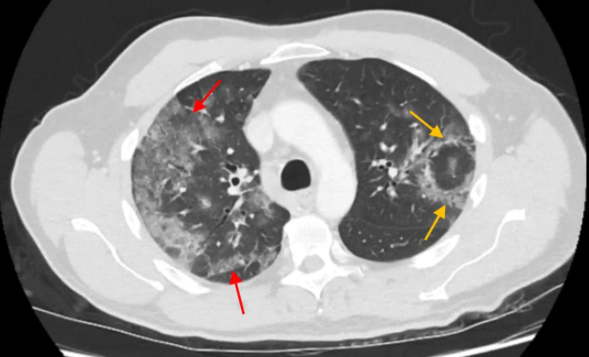
Reversed halo sign is caused by a focal process resulting in central ground-glass opacity surrounded by a more dense ring of consolidation in the lung parenchyma. This CT imaging finding is most characteristically seen in cryptogenic organizing pneumonia (COP) but also occurs in a variety of infectious and noninfectious underlying pathologies, including invasive fungal infections (e.g., pulmonary mucormycosis, aspergillosis), Pneumocystis jirovecii pneumonia, tuberculosis, pulmonary infarction, granulomatous vasculitis (formerly Wegener’s granulomatosis), lymphomatoid granulomatosis, sarcoidosis, and even in COVID-19 pneumonia. This sign reflects an organizing inflammatory or infectious process with variable involvement of alveolar spaces and interstitium.
Why is it called so?
The name reversed halo sign refers to the characteristic imaging appearance on CT where a central area of ground-glass opacity is encircled by a denser consolidation, forming a ring-like shape opposite to the classical “halo sign,” where a denser nodule is surrounded by ground-glass opacity. Hence, it is a “reversed” pattern compared to the conventional halo.
Pathophysiology
The central ground-glass opacity corresponds to alveolar septal inflammation and partial filling of alveolar spaces with inflammatory exudate, maintaining some air content. The surrounding denser consolidation represents more complete alveolar filling with organizing pneumonia (fibroblastic tissue plugging in alveolar ducts and alveoli) or peripheral granulomatous inflammation. The ring of consolidation forms due to peripheral extension of inflammatory cells and fibrosis around the central area, creating the distinctive reversed halo sign pattern.
Alternative names: None commonly used
Other associated named signs:
- Halo sign (related but opposite pattern)
- May coexist with additional CT findings such as centrilobular nodules in tuberculosis or cavitation in fungal infections
Access all radiology signs posted so far: https://radiogyan.com/radiology-signs/

Improve Content And Help Your Colleagues!
Found an error in the post? Please let us know using our contact page and we will update it with due credits!
If you wish to contribute radiological images for this case (or any other article on the website), you can submit them here and you will be duly credited: Submit radiology case