


Key points
- The WHO-IWGE classification stages hydatid cysts on ultrasound into CL (undifferentiated), active CE1 and CE2, transitional CE3a and CE3b, and inactive CE4 and CE5 [1,2].
- It is an ultrasound classification. MRI reproduces it well (kappa 0.83 to 1.0 for CE1 to CE4); CT does not (kappa 0.62 to 0.72), except for CE5 [6].
- CE3a and CE3b behave differently. CE3a (water lily sign) responds like CE1; CE3b (daughter cysts in a solid matrix) behaves like CE2 and is the least responsive stage to non-surgical treatment [2,12].
- Treatment is stage-specific: albendazole or PAIR for CE1 and CE3a, catheterization or surgery for CE2 and CE3b, watch and wait for uncomplicated CE4 and CE5 [3,10].
- Calcification is not the same as CE5. Calcium appears at every stage; 26 of 67 calcified cysts in one series were still CE1 to CE3 by content [7].
- Uncomplicated inactive liver cysts stay inactive: 98.5 percent over long-term follow-up without treatment [13].
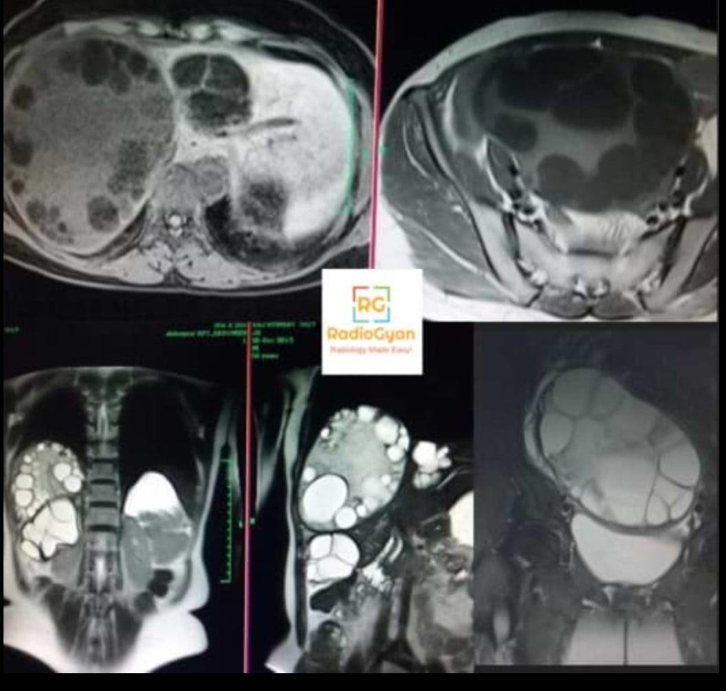
Cystic echinococcosis (hydatid disease) is caused by the larval stage of Echinococcus granulosus sensu lato, and the liver is the commonest site [14]. The cyst evolves over years from a fluid-filled, fertile lesion to a solid, calcified, dead one, and what to do with it depends almost entirely on where it sits along that path. That is why staging matters more than detection: the stage decides between drugs, a needle, a surgeon or nothing at all [3,5].
The WHO Informal Working Group on Echinococcosis (WHO-IWGE) standardised ultrasound staging in 2003 [1]. The original CE3 stage was later divided into CE3a and CE3b, because cysts with a detached membrane and cysts with daughter cysts in a solid matrix respond to treatment very differently [2].
WHO-IWGE classification at a glance
| Stage | Activity | Ultrasound appearance | Named sign | Gharbi type |
|---|---|---|---|---|
| CL | Undifferentiated | Unilocular anechoic cyst, no visible wall | None | Not classified |
| CE1 | Active, usually fertile | Unilocular anechoic cyst with a visible double-line wall; may contain floating echogenic foci | Double line sign, snowflake sign (hydatid sand) | I |
| CE2 | Active, usually fertile | Multivesicular, multiseptated cyst; daughter cysts fill the cavity or line a mother cyst | Honeycomb, rosette or wheel-spoke pattern | III |
| CE3a | Transitional | Cyst with a detached laminated membrane floating in the fluid | Water lily sign | II |
| CE3b | Transitional, often still active | Daughter cysts within a solid, echogenic matrix | Daughter cysts in a solid matrix | III |
| CE4 | Inactive | Heterogeneous solid content, no daughter cysts; folded membranes | Ball of wool (ball of yarn) | IV |
| CE5 | Inactive | Solid content with a thick, partly or completely calcified wall and posterior acoustic shadowing | Arch-like or eggshell calcification | V |
Stage-by-stage imaging features
CL: undifferentiated cystic lesion
A unilocular, anechoic cyst with no visible parasitic wall. It is indistinguishable from a simple hepatic cyst on morphology alone. If it is echinococcal, it is usually an early, not yet fertile lesion [1,3]. Do not call it a hydatid cyst on imaging. Look hard for a double wall with a high-frequency probe, check serology, and re-image: the appearance of a wall converts it to CE1 [5].
CE1: unilocular cyst with a visible wall
A round, anechoic cyst with a double-line wall: the echogenic laminated layer of the parasite against the host pericyst. Fine echogenic foci (hydatid sand, made of brood capsules and protoscolices) may settle dependently and swirl when the patient turns, the snowflake sign [1,14]. CE1 is active and usually fertile.
CE2: multivesicular cyst with daughter cysts
The cyst is divided by septa into multiple compartments: daughter cysts budding from the germinal layer. They may pack the whole cavity (honeycomb) or line the periphery around a central mother cyst (rosette, wheel-spoke) [1,14]. CE2 is active and fertile, and its septated structure is the reason PAIR fails: the sclerosant cannot reach every compartment [10].
CE3a: detached membrane (water lily sign)
Loss of intracystic pressure lets the laminated membrane separate from the pericyst. A partial split gives a double contour along one wall; a complete collapse leaves the undulating membrane floating in the fluid, the water lily sign [1]. CE3a is the start of degeneration. It behaves like CE1 for treatment purposes [3].
CE3b: daughter cysts in a solid matrix
Daughter cysts persist inside an echogenic, solid-looking matrix of degenerated membranes and debris. The matrix makes it look half-dead, but it is not: CE3b is the least responsive stage to non-surgical treatment and relapses frequently [2,12]. In a Pavia series of 60 patients, albendazole produced only temporary inactivation of CE3b, while untreated cysts under watch and wait stayed stable with no difference in complications [12].
CE4: heterogeneous solid content
The cyst has lost its fluid. The content is heterogeneous, hypo- or hyperechoic, with tightly folded membranes giving the ball of wool appearance, and no daughter cysts [1,14]. CE4 is the stage most often mistaken for a tumour; its avascular content and the folded-membrane pattern are the clues.
CE5: calcified wall
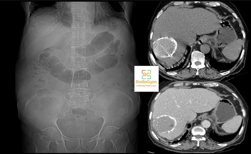
Solid content with a thick calcified wall, arch-like on ultrasound, producing a posterior acoustic shadow that may hide the content entirely [1]. CE5 is the end stage and is inactive, but only when the content is solid as well. A calcified wall around fluid or daughter cysts is not CE5 [7].
Natural history: why the order matters
The WHO-IWGE stages follow the life of the cyst. The usual sequence runs from CE1 through CE3a to CE4 and CE5, or from CE1 to CE2, CE3b and then CE4 [2,14]. The path is not one-way. Cysts made inactive by albendazole can revert: in a pooled analysis of 1,159 liver and peritoneal cysts, about 25 percent of responders reverted to an active stage within 1.5 to 2 years [9]. A CE4 cyst after drug treatment is therefore less reliably dead than a CE4 cyst found untreated.
Gharbi vs WHO-IWGE classification
| Gharbi (1981) | Description | WHO-IWGE |
|---|---|---|
| Not classified | Cyst without a visible wall | CL |
| Type I | Pure fluid collection | CE1 |
| Type II | Fluid collection with a split wall | CE3a |
| Type III | Fluid collection with septa or daughter cysts | CE2, or CE3b when the matrix is solid |
| Type IV | Heterogeneous echo pattern | CE4 |
| Type V | Reflecting thick walls | CE5 |
Gharbi described the patterns first, but its numbering puts the detached membrane (type II) before daughter cysts (type III), which does not match how cysts evolve. It also has no place for undifferentiated lesions. The WHO-IWGE scheme fixed both and groups the stages by biological activity, which is what treatment decisions need [1,2].
Staging on CT and MRI
- Ultrasound stays the reference. The classification was defined on ultrasound, and ultrasound is the method of choice for staging and follow-up wherever the organ is accessible [6,5].
- MRI is the best substitute. In 187 abdominal cysts, MRI agreed with ultrasound with kappa 0.83 to 1.0 for CE1 to CE4. Heavily T2-weighted sequences show the membranes, daughter cysts and matrix best [6].
- CT is weak for CE1 to CE4 (kappa 0.62 to 0.72) because it shows the fine internal membranes poorly, but it is the best tool for CE5 (kappa 0.95) [6].
- Calcification on CT does not stage the cyst. Half of all cysts in a Heidelberg series were calcified, and over a third of those were still CE1 to CE3. Stage by the content, not the wall [7].
- MRI matrix signs of inactivity. A 2026 multicentre study of 174 cysts found fatty T1 hyperintensity and a “matrix flip-flop” sign each highly specific for inactivity (97.4 percent), and matrix calcification strongly associated with inactive stages [8].
Stage-specific management
| Stage | First-line approach | Notes |
|---|---|---|
| CL | Further characterisation | Serology, high-resolution ultrasound or MRI, and follow-up imaging before any treatment |
| CE1, CE3a under 5 cm | Albendazole alone | CE1 responds best to benzimidazoles: 50 to 75 percent inactive or gone at 1 to 2 years [9] |
| CE1, CE3a over 5 cm | PAIR with albendazole cover | Standard catheterization if a cysto-biliary fistula appears or PAIR is technically difficult [10] |
| CE2, CE3b | Modified catheterization (MoCaT), surgery, or albendazole | Drug response only 30 to 55 percent [9]. MoCaT: 4.5 percent recurrence over a median of 49 months in 132 cysts [11]. Asymptomatic CE3b may be watched [12] |
| CE4, CE5 uncomplicated | Watch and wait | 98.5 percent of untreated inactive liver cysts stayed inactive on long-term follow-up [13] |
| Any stage complicated | Surgery or intervention | Rupture, cysto-biliary fistula, infection or mass effect override the stage |
What to put in the report
- The WHO-IWGE stage of each cyst, not just “hydatid cyst”. The stage is the treatment decision.
- Size in three planes and location, with the Couinaud segment for liver cysts and the relation to the hepatic hilum, major vessels and bile ducts.
- Number of cysts, and whether they are at different stages. Mixed stages are common.
- Wall and content: double line, detached membrane, daughter cysts, matrix, calcification. If calcified, say whether the content is fluid or solid.
- Complications: biliary communication (bile duct dilatation, fat-fluid level, membranes in the ducts), rupture, infection (gas, air-fluid level, wall enhancement), mass effect.
- Extrahepatic disease: peritoneal, splenic and pulmonary cysts change management.
- Comparison with prior imaging, especially after treatment, since reversion from an inactive to an active stage signals relapse.
Pitfalls
- Calling a CL a hydatid cyst. Without a visible wall, a unilocular cyst is a simple cyst until proven otherwise.
- Calling CE3b inactive. The solid matrix looks degenerate, but daughter cysts inside it mean the cyst is still capable of activity [2].
- Staging on CT alone. CT under-shows membranes and daughter cysts; use ultrasound or T2-weighted MRI [6].
- Equating calcification with death. Stage by the content [7].
- Mistaking CE4 for a tumour. A solid-looking, avascular liver mass with layered or folded internal lines in a patient from an endemic area should raise CE4 before biopsy.
Related reading on RadioGyan
- Water lily sign: the detached membrane of CE3a.
- Hepatic cystic lesions: the differential of a cystic liver lesion.
- Caroli disease: another cystic liver mimic, with the central dot sign.
Frequently asked questions
References
- WHO Informal Working Group on Echinococcosis. International classification of ultrasound images in cystic echinococcosis for application in clinical and field epidemiological settings. Acta Trop. 2003;85(2):253-261. PMID: 12606104.
- Junghanss T, da Silva AM, Horton J, Chiodini PL, Brunetti E. Clinical management of cystic echinococcosis: state of the art, problems, and perspectives. Am J Trop Med Hyg. 2008;79(3):301-311. PMID: 18784219.
- Brunetti E, Kern P, Vuitton DA; Writing Panel for the WHO-IWGE. Expert consensus for the diagnosis and treatment of cystic and alveolar echinococcosis in humans. Acta Trop. 2010;114(1):1-16. PMID: 19931502.
- Gharbi HA, Hassine W, Brauner MW, Dupuch K. Ultrasound examination of the hydatic liver. Radiology. 1981;139(2):459-463. PMID: 7220891.
- Tamarozzi F. Ultrasound diagnosis of cystic echinococcosis: updates and implications for clinical management. J Helminthol. 2024;98:e34. PMID: 38628145.
- Stojkovic M, Rosenberger K, Kauczor HU, Junghanss T, Hosch W. Diagnosing and staging of cystic echinococcosis: how do CT and MRI perform in comparison to ultrasound? PLoS Negl Trop Dis. 2012;6(10):e1880. PMID: 23145199.
- Hosch W, Stojkovic M, Janisch T, Kauffmann GW, Junghanss T. The role of calcification for staging cystic echinococcosis (CE). Eur Radiol. 2007;17(10):2538-2545. PMID: 17473925.
- Julien M, Bohard L, Busse-Cote A, Tissot N, Turco C, Barerra C, et al. From fluid loss to calcification: MRI and CT matrix features in cystic echinococcosis. Insights Imaging. 2026;17(1):218. PMID: 42690489.
- Stojkovic M, Zwahlen M, Teggi A, Vutova K, Cretu CM, Virdone R, et al. Treatment response of cystic echinococcosis to benzimidazoles: a systematic review. PLoS Negl Trop Dis. 2009;3(9):e524. PMID: 19787039.
- Akhan O. Percutaneous treatment of liver hydatid cysts: to PAIR or not to PAIR. Curr Opin Infect Dis. 2023;36(5):308-317. PMID: 37548385.
- Akhan O, Ozbay Y, Unal E, Karaagaoglu E, Ciftci TT, Akinci D. Long-term results of modified catheterization technique in the treatment of CE type 2 and 3b liver hydatid cysts. Cardiovasc Intervent Radiol. 2025;48(4):503-511. PMID: 39953155.
- Rinaldi F, De Silvestri A, Tamarozzi F, Cattaneo F, Lissandrin R, Brunetti E. Medical treatment versus “Watch and Wait” in the clinical management of CE3b echinococcal cysts of the liver. BMC Infect Dis. 2014;14:492. PMID: 25204575.
- Lissandrin R, Tamarozzi F, Mariconti M, Manciulli T, Brunetti E, Vola A. Watch and wait approach for inactive echinococcal cyst of the liver: an update. Am J Trop Med Hyg. 2018;99(2):375-379. PMID: 29869600.
- Rinaldi F, Brunetti E, Neumayr A, Maestri M, Goblirsch S, Tamarozzi F. Cystic echinococcosis of the liver: a primer for hepatologists. World J Hepatol. 2014;6(5):293-305. PMID: 24868323.

Improve Content And Help Your Colleagues!
Found an error in the post? Please let us know using our contact page and we will update it with due credits!
If you wish to contribute radiological images for this case (or any other article on the website), you can submit them here and you will be duly credited: Submit radiology case