


Key facts for practice and radiology board exams
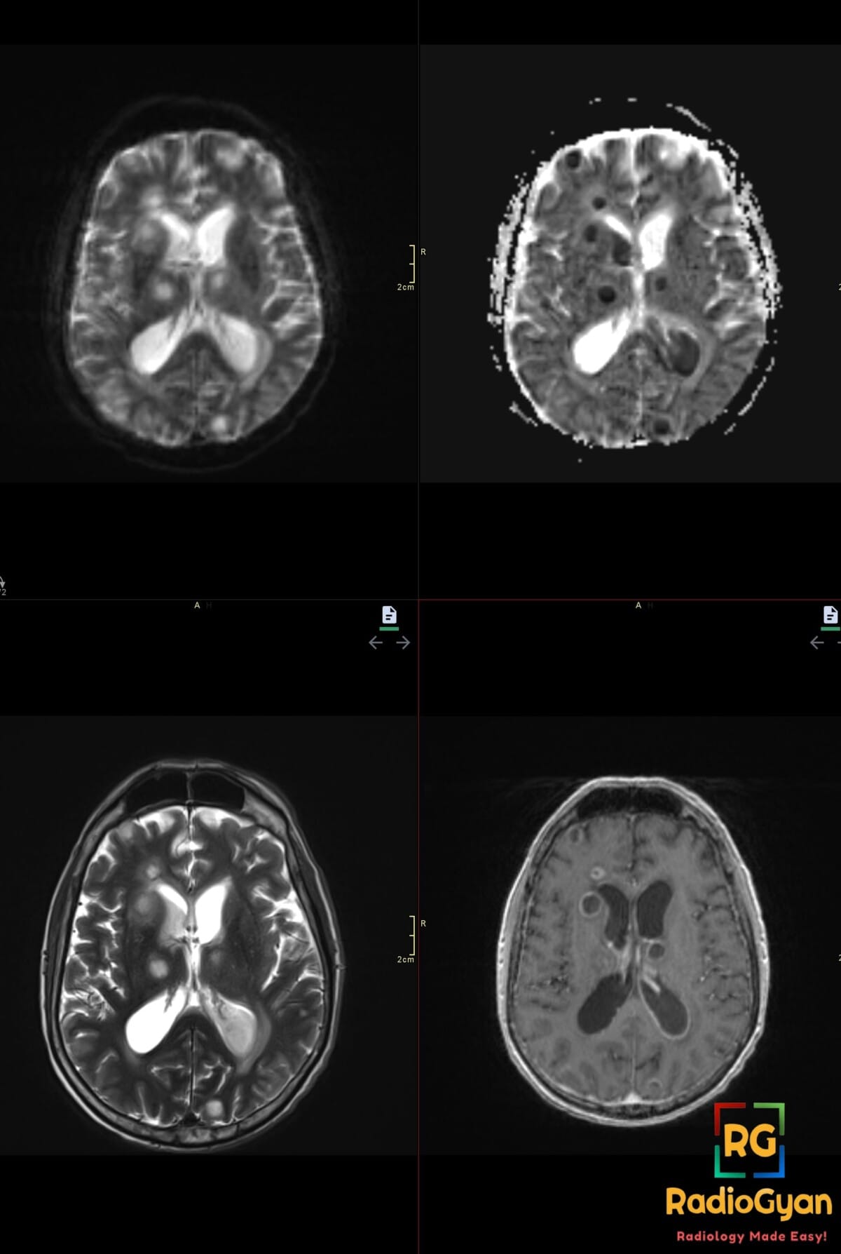
A ring enhancing lesion in the brain is a rim of enhancement around a non-enhancing centre. The eleven causes worth holding in mind are abscess, tuberculoma, neurocysticercosis, toxoplasmosis, lymphoma, glioblastoma, metastasis, tumefactive demyelination, subacute infarct, radiation necrosis and resolving haematoma. Diffusion settles most of it: a centre that restricts diffusion with a low ADC is pus, not tumour.
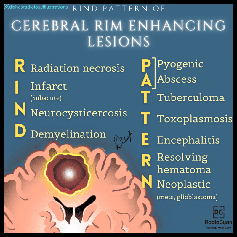
RIND PATTERN mnemonic for ring enhancing brain lesions
Ring enhancing lesions follow a RIND PATTERN of enhancement.
R – Radiation necrosis
I – Infarct (subacute)
N – Neurocysticercosis
D – Demyelination (tumefactive)
P – Pyogenic
A – Abscess
T – Tuberculoma
T – Toxoplasmosis
E – Encephalitis
R – Resolving haematoma
N – Neoplastic (metastases, glioblastoma, lymphoma)
The shorter version is MAGIC DR, also written MAGIC Lady Doctor or MAGICAL DR when lymphoma is included.
M – Metastasis
A – Abscess
G – Glioblastoma
I – Infarct (subacute phase)
C – Contusion
A/Lady – Lymphoma
D – Demyelination (tumefactive)
R – Radiation necrosis
How to tell ring enhancing lesions apart on MRI
The mnemonic gives eleven names, not an answer. ADC is quoted in 10-3 mm2/s, rCBV as a ratio to contralateral white matter.
| Cause | Ring morphology | Centre on DWI | The finding that narrows it |
|---|---|---|---|
| Pyogenic abscess | Smooth, thin, thinner on the ventricular side | Restricted, ADC about 0.65 | Dual rim sign on SWI; T2 hypointense rim |
| Tuberculoma | Thick, often conglomerate rings | Variable | T2 hypointense centre; ADC separates it from glioma better than choline does |
| Neurocysticercosis | Thin, around a cyst under 2 cm | Not restricted | Scolex as a mural dot in a hole |
| Toxoplasmosis | Basal ganglia and grey-white junction | Not restricted | Eccentric or concentric target sign; rCBV ratio about 0.84 |
| Primary CNS lymphoma | Rings mainly in immunosuppression; solid if immunocompetent | Restricted in the solid part | Periventricular with subependymal spread; shrinks on steroids |
| Glioblastoma | Thick, irregular, nodular, crosses the corpus callosum | Not restricted | rCBV ratio about 4.5; tumour extends beyond the enhancing rim |
| Metastases | Multiple, thin, at the grey-white junction | Not restricted | rCBV ratio about 3.6; oedema out of proportion to size |
| Tumefactive demyelination | Incomplete ring confined to the white matter | Not restricted | Open ring sign; little mass effect for the size |
| Subacute infarct | Gyriform, within one arterial territory | Pseudonormalising by this stage | Parenchymal enhancement is a late finding, from the second week |
| Radiation necrosis | Patchy, inside the radiation port | Not restricted | Lower rCBV than recurrent tumour; no new solid nodule |
| Resolving haematoma | Thin, smooth, complete | Not restricted | Blood products on SWI and GRE; shrinks on follow-up |
Five named signs that settle most cases
| Sign | What you see | What it means |
|---|---|---|
| Open ring sign | Incomplete enhancing ring or crescent confined to the white matter | Atypical demyelination, specificity 94 percent and 84 percent for two readers |
| Dual rim sign | Two concentric rims on SWI, outer hypointense and inner hyperintense | Pyogenic abscess: present in 9 of 12 abscesses and 0 of 20 glioblastomas |
| Eccentric target sign | Enhancing nodule on one wall of the ring | Cerebral toxoplasmosis, the nodule being inflamed vessels along a sulcus |
| Concentric target sign | Alternating T2 rings on non-enhanced images | Also toxoplasmosis, and thought to be the more specific of the two |
| Dot in a hole | Eccentric mural dot inside a small cyst | Scolex of a degenerating cysticercus, one of three absolute criteria |
Both numbers have exceptions. Abscesses with ADC outside the usual range appear in every series, and a treated or very early abscess can behave like a tumour. An open ring is highly specific, not diagnostic: neoplasm and infection remain frequent causes of incomplete rim enhancement.
Why the ring enhances
Enhancement marks where the blood brain barrier has failed, so the ring is granulation tissue or viable tumour, not the edge of the lesion. In an abscess it is an organised collagen capsule: smooth, T2 hypointense, and thinner on the ventricular side where it matures last. In a glioblastoma it is cellular tumour with neovascularity, so it is thick, irregular and high on perfusion, and tumour extends well beyond it into the T2 hyperintense oedema. That one difference explains the imaging, and why the biopsy that yields tissue is aimed at the ring rather than the centre.
Working through a ring enhancing lesion
- Read the centre before the ring. Restricted diffusion with a low ADC means pus, or less often the solid part of a lymphoma.
- Get susceptibility weighted imaging: it carries the dual rim sign, the blood products of a resolving haematoma and the vessels of a tumour rim.
- An incomplete ring confined to the white matter is demyelination until proven otherwise. Add perfusion for tumour versus not tumour, and for recurrence versus radiation necrosis in a treated field.
- Anchor on the setting. Immune status, endemic exposure, a known primary, recent radiotherapy and time since onset reorder the list more than any single MRI feature.
A naming point worth keeping current: under the 2021 WHO classification, glioblastoma is IDH-wildtype by definition and the term glioblastoma multiforme is retired. An identical looking ring that proves IDH-mutant is astrocytoma, IDH-mutant, grade 4.
Frequently asked questions
References
- Toh CH, Wei KC, Chang CN, Hsu PW, Wong HF, Ng SH, et al. Differentiation of pyogenic brain abscesses from necrotic glioblastomas with use of susceptibility-weighted imaging. AJNR Am J Neuroradiol. 2012;33(8):1534-8. PMID: 22422181.
- Chang SC, Lai PH, Chen WL, Weng HH, Ho JT, Wang JS, et al. Diffusion-weighted MRI features of brain abscess and cystic or necrotic brain tumors: comparison with conventional MRI. Clin Imaging. 2002;26(4):227-36. PMID: 12140151.
- Masdeu JC, Quinto C, Olivera C, Tenner M, Leslie D, Visintainer P. Open-ring imaging sign: highly specific for atypical brain demyelination. Neurology. 2000;54(7):1427-33. PMID: 10751251.
- Mahadevan A, Ramalingaiah AH, Parthasarathy S, Nath A, Ranga U, Krishna SS. Neuropathological correlate of the concentric target sign in MRI of HIV-associated cerebral toxoplasmosis. J Magn Reson Imaging. 2013;38(2):488-95. PMID: 23440973.
- Del Brutto OH, Nash TE, White AC Jr, Rajshekhar V, Wilkins PP, Singh G, et al. Revised diagnostic criteria for neurocysticercosis. J Neurol Sci. 2017;372:202-10. PMID: 28017213.
- Muccio CF, Esposito G, Bartolini A, Cerase A. Cerebral abscesses and necrotic cerebral tumours: differential diagnosis by perfusion-weighted magnetic resonance imaging. Radiol Med. 2008;113(5):747-57. PMID: 18414811.
- Peng J, Ouyang Y, Fang WD, Luo TY, Li YM, Lv FJ, et al. Differentiation of intracranial tuberculomas and high grade gliomas using proton MR spectroscopy and diffusion MR imaging. Eur J Radiol. 2012;81(12):4057-63. PMID: 22749802.
- Barajas RF Jr, Chang JS, Segal MR, Parsa AT, McDermott MW, Berger MS, et al. Differentiation of recurrent glioblastoma multiforme from radiation necrosis after external beam radiation therapy with dynamic susceptibility-weighted contrast-enhanced perfusion MR imaging. Radiology. 2009;253(2):486-96. PMID: 19789240.
- Louis DN, Perry A, Wesseling P, Brat DJ, Cree IA, Figarella-Branger D, et al. The 2021 WHO classification of tumors of the central nervous system: a summary. Neuro Oncol. 2021;23(8):1231-51. PMID: 34185076.
- Elster AD, Moody DM. Early cerebral infarction: gadopentetate dimeglumine enhancement. Radiology. 1990;177(3):627-32. PMID: 2243961.
Browse more high-yield revision aids on our radiology mnemonics collection.
Check out more visual mnemonics here:
Ankle Tendons Anatomy- Visual Mnemonics
Neurofibromastosis 2 – Visual Mnemonics

Hyperdense Lesions On NCCT – Visual Mnemonics

Improve Content And Help Your Colleagues!
Found an error in the post? Please let us know using our contact page and we will update it with due credits!
If you wish to contribute radiological images for this case (or any other article on the website), you can submit them here and you will be duly credited: Submit radiology case
Thanks a lot for this important radiological information
Glad you like it, Rasha! You can check out more illustrated mnemonics here: Radiology Visual Mnemonics