
Primary Sclerosing Cholangitis – Radiology Board Review
- Clinical: Middle-aged men (peak 30-50 years); strong association with inflammatory bowel disease (particularly ulcerative colitis); progressive fatigue, pruritus, jaundice; increased risk of cholangiocarcinoma and colorectal cancer; may be asymptomatic with only elevated alkaline phosphatase
- Etiology/Pathophys: Chronic immune-mediated cholangiopathy causing progressive inflammatory fibrosis of intra- and extrahepatic bile ducts; leads to biliary strictures, cholestasis, and eventual cirrhosis; gut-liver axis involvement with altered microbiome
- Radiograph: Generally normal in early stages; may show hepatomegaly or features of portal hypertension in advanced disease
- US: Dilated intrahepatic bile ducts with irregular walls; echogenic portal tract thickening; hepatomegaly; splenomegaly in advanced cases; may show gallbladder wall thickening
- CT: Alternating strictures and dilatations of bile ducts creating “beading” appearance; wall thickening of bile ducts; hepatomegaly; portal lymphadenopathy; signs of cirrhosis and portal hypertension in advanced disease
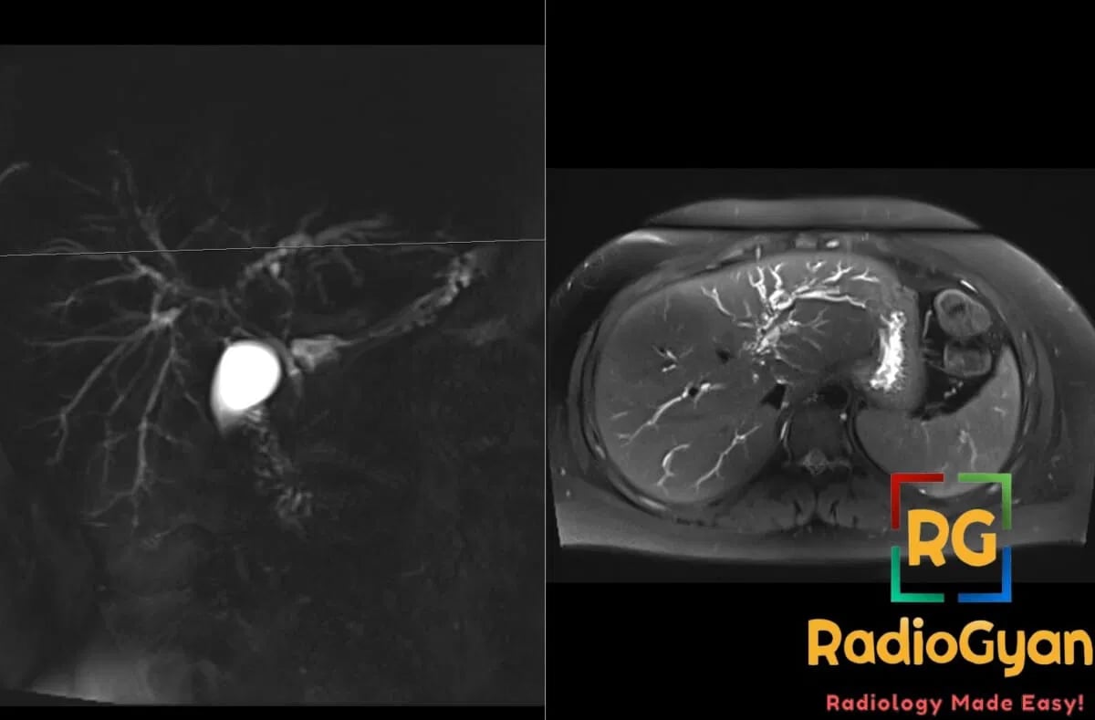
- MRI: MRCP is diagnostic modality of choice; classic “string of beads” sign with alternating strictures and dilatations; pruned tree appearance of intrahepatic ducts; T2 hyperintense periductal thickening; dominant strictures appear as severe focal narrowing
- Signs: String of beads sign – alternating strictures and dilatations on MRCP; pruned tree sign – loss of peripheral bile duct branching; periductal halo sign – T2 hyperintense rim around bile ducts
- Frameworks: Classification based on involvement: large duct PSC (classic), small duct PSC, PSC-autoimmune hepatitis overlap syndrome; staging based on extent of ductal involvement and presence of cirrhosis
- DDx: Secondary sclerosing cholangitis (ischemic, infectious, toxic); cholangiocarcinoma (focal stricture, mass lesion); recurrent pyogenic cholangitis (stones, abscesses); IgG4-related sclerosing cholangitis (pancreatic involvement, elevated IgG4)
- Tx: UDCA for symptom management; endoscopic therapy for dominant strictures; liver transplantation for end-stage disease; radiologist monitors for cholangiocarcinoma development and disease progression

Improve Content And Help Your Colleagues!
Found an error in the post? Please let us know using our contact page and we will update it with due credits!
If you wish to contribute radiological images for this case (or any other article on the website), you can submit them here and you will be duly credited: Submit radiology case