
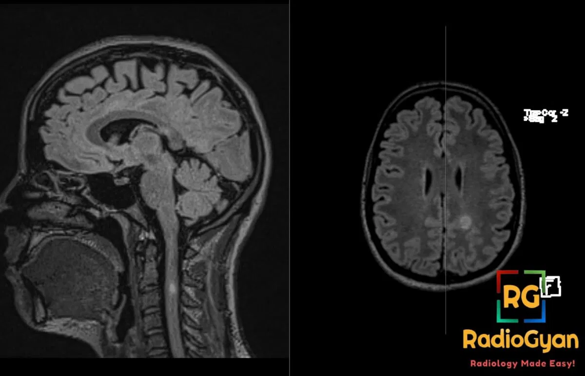
32 yr female presenting with subacute motor weakness and sensory disturbances.
Diagnosis and teaching points:
Diagnosis: Multiple sclerosis
Teaching points:
- Clinical: Typically affects young adults (20-40 years); 3:1 female predominance; presents with relapsing-remitting neurological deficits like optic neuritis, motor weakness, or sensory disturbances; risk factors include HLA-DR2, low vitamin D, smoking, and EBV infection.
- Etiology/Pathophys: Immune-mediated demyelinating disease where autoreactive T-cells cause multifocal inflammation, axonal loss, and gliosis in the central nervous system white matter.
- CT: Low sensitivity; may show non-specific hypodense white matter lesions.
- MRI: T2/FLAIR hyperintense lesions in periventricular, juxtacortical, infratentorial, and spinal cord regions; chronic lesions appear as T1 hypointense “black holes”; active lesions show variable gadolinium enhancement; spinal lesions are typically short-segment (less than 2 vertebral levels).
- Signs: Dawson’s fingers (ovoid lesions perpendicular to ventricles); Open-ring enhancement (incomplete enhancement favoring demyelination over tumor); Central vein sign (vein within lesion on susceptibility-weighted imaging); Black holes (axonal loss).
- Frameworks: 2017 McDonald criteria require dissemination in space (lesions in 2+ characteristic regions) and time (combination of enhancing/non-enhancing lesions, new lesions on follow-up, or CSF oligoclonal bands).
- DDx: ADEM (monophasic, deep gray matter involvement); Small vessel ischemia (older patients, lacks perpendicular orientation or spinal lesions); PML (confluent U-fiber involvement, microcysts); Glioma (mass effect, complete ring enhancement).
- Tx: Disease-modifying therapies (interferons, ocrelizumab, natalizumab) to reduce relapses; high-dose corticosteroids for acute exacerbations.
OSCE Questions
Question: What is the classic name for the ovoid white matter lesions oriented perpendicular to the ventricles along the medullary veins?
Dawson’s fingers.
Question: Which clinical criteria are standard for establishing the diagnosis based on dissemination in space and time?
2017 McDonald criteria.
Question: What pattern of gadolinium enhancement is highly suggestive of this demyelinating pathology over a neoplastic process?
Open-ring enhancement.
Question: Presence of what specific finding on susceptibility-weighted imaging (SWI) helps distinguish this condition from small vessel ischemic disease?
Central vein sign.
Question: What do persistent T1 hypointense “black holes” signify regarding the underlying neural tissue?
Permanent axonal loss.
MCQ Questions
1. What is the classic orientation of these lesions relative to the lateral ventricles?
A. Parallel to ventricles
B. Perpendicular to ventricles
C. Circumferential to ventricles
D. Randomly scattered
Answer: B. Perpendicular to ventricles. Known as Dawson’s fingers, these ovoid lesions follow the orientation of medullary veins.
2. Which MRI finding is most specific for this demyelinating pathology over a tumor?
A. Complete ring enhancement
B. Mass effect
C. Open-ring enhancement
D. Restricted diffusion
Answer: C. Open-ring enhancement. An incomplete ring of enhancement is highly suggestive of demyelination rather than a neoplastic process.
3. What does a “black hole” on T1-weighted sequences signify in this condition?
A. Active inflammation
B. Acute hemorrhage
C. Permanent axonal loss
D. Calcification
Answer: C. Permanent axonal loss. Persistent T1 hypointensity represents areas of irreversible tissue matrix destruction and axonal loss.
4. Which location is part of the McDonald criteria for dissemination in space?
A. Thalamus
B. Optic nerve
C. Infratentorial white matter
D. Internal capsule
Answer: C. Infratentorial white matter. The four characteristic regions include periventricular, cortical/juxtacortical, infratentorial, and spinal cord.
5. What feature on susceptibility-weighted imaging helps distinguish this from small vessel ischemia?
A. Cortical sparing
B. Central vein sign
C. Lacunar infarcts
D. Microbleeds
Answer: B. Central vein sign. The presence of a small vessel in the center of a white matter lesion is highly specific for this disease.
Check out more such cases:

Improve Content And Help Your Colleagues!
Found an error in the post? Please let us know using our contact page and we will update it with due credits!
If you wish to contribute radiological images for this case (or any other article on the website), you can submit them here and you will be duly credited: Submit radiology case