
65 yr male with a groin mass following a catheterization procedure.
Diagnosis and teaching points:
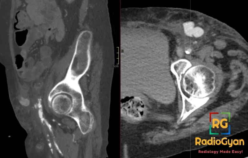
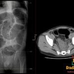
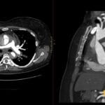
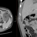
Diagnosis: Femoral artery pseudoaneurysm post femoral access for coronary angiogram.jpg
Teaching points:
- Clinical: Incidence 0.05-14% post-catheterization (e.g., coronary angiogram via femoral access); symptoms: pulsatile hematoma, pain, ecchymosis, active extravasation; risk factors: female gender, anticoagulation, large sheath size, obesity, hemodialysis, poor technique, PAD, elderly, low/high puncture sites, therapeutic interventions; etymology: “pseudo” (false) aneurysm, involves outpouching of only 1-2 vessel wall layers.
- Etiology/Pathophys: Arterial wall disruption from percutaneous puncture (e.g., femoral access), leading to blood extravasation contained by surrounding hematoma/soft tissues, forming a saccular outpouching connected to the artery by a narrow neck; blood enters in systole, exits in diastole.
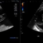
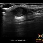
- US: First-line (92-97% sensitivity/specificity) shows saccular collection with narrow neck, “to-and-fro” or “yin-yang” flow (systolic filling/red-blue swirling on color Doppler, diastolic emptying on spectral Doppler), thrombus, surrounding hematoma; operator-dependent, limited in deep vessels or trauma.
- CT: Contrast-enhanced CT/CTA detects saccular pseudoaneurysm as rounded/ovoid contrast-filled collection adjacent to artery with narrow neck/stalk; unenhanced shows hematoma-like mass; assesses extent, surrounding induration, atherosclerosis, and vessels.
- Signs: To-and-fro sign: Spectral Doppler shows forward systolic flow into sac and reverse diastolic flow out; Yin-yang sign: Color Doppler swirling red-blue flow in sac due to bidirectional swirling.
- DDx: Hematoma: No flow/neck on US/CT, lacks to-and-fro/yin-yang, uniform on imaging; True aneurysm: All 3 vessel wall layers, fusiform, no narrow neck; AV fistula: High-velocity low-resistance flow, direct artery-vein shunt without discrete sac; Abscess/cellulitis: No vascular flow/contrast enhancement in sac, clinical fever/infection.
- Tx: Ultrasound-guided compression (first-line for small/stable); thrombin injection; endovascular (covered stent/coil embolization); surgery for large/failing cases; monitor with serial US.
OSCE Questions
Question: What is the most common etiology for this pathology?
Percutaneous arterial puncture, often after catheterization procedures.
Question: What specific Doppler ultrasound finding is considered the “hallmark” for diagnosing this condition?
The “to-and-fro” flow pattern on spectral Doppler.
Question: What imaging modality is considered first-line for evaluating this pathology?
Duplex ultrasound (DUS).
Question: Name one other condition that could be in the differential diagnosis for this pathology.
Hematoma, true aneurysm, or AV fistula are all viable answers.
Question: What is a common non-surgical treatment approach for small, stable cases of this pathology?
Ultrasound-guided compression.[/spoler]
MCQ Questions
1. What imaging modality is first-line for this diagnosis?
A. CT
B. MRI
C. Ultrasound
D. Radiography
Answer: C. Ultrasound. Duplex ultrasound is the first-line imaging modality for diagnosing femoral artery pseudoaneurysms.
2. What color Doppler sign is characteristic of this pathology?
A. Target sign
B. Yin-yang sign
C. Pseudokidney sign
D. Double-rim sign
Answer: B. Yin-yang sign. The yin-yang sign describes the swirling red-blue flow seen on color Doppler in a pseudoaneurysm sac.
3. What spectral Doppler finding is characteristic of this pathology?
A. High-resistance triphasic flow
B. To-and-fro flow
C. Monophasic flow
D. Reversed diastolic flow
Answer: B. To-and-fro flow. The to-and-fro sign, showing forward systolic and reverse diastolic flow, is a hallmark on spectral Doppler.
4. Which of these is a risk factor for developing this pathology?
A. Small sheath size
B. Anticoagulation
C. Low BMI
D. Diagnostic intervention
Answer: B. Anticoagulation. Anticoagulation is a significant risk factor for pseudoaneurysm formation after arterial puncture.
5. This pathology’s outpouching involves how many vessel wall layers?
A. All three
B. One to two
C. Zero
D. Three to four
Answer: B. One to two. A pseudoaneurysm is distinct from a true aneurysm as it involves only one to two layers of the arterial wall, not all three.
Check out more such cases:

Improve Content And Help Your Colleagues!
Found an error in the post? Please let us know using our contact page and we will update it with due credits!
If you wish to contribute radiological images for this case (or any other article on the website), you can submit them here and you will be duly credited: Submit radiology case