
What causes gloved finger sign in the lungs on chest radiography or CT?
Let me know in the comments.
Click to reveal the answer
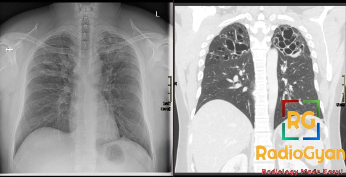
Gloved finger sign is caused by mucoid impaction within dilated bronchi, appearing as branching, finger-like tubular opacities radiating from the hilum on chest radiography or CT. This imaging finding is commonly associated with conditions that cause airway obstruction or bronchial dilation, including allergic bronchopulmonary aspergillosis (ABPA), bronchial atresia, bronchiectasis, cystic fibrosis, broncholithiasis, bronchial tuberculosis, and foreign body aspiration. Clinically, these conditions involve mucus plugging due to impaired clearance or direct bronchial obstruction.
Why is it called so?
The sign is named “gloved finger” because the radiologic branching, finger-like opacities resemble the shape of a finger of a glove; the mucus filling dilated bronchi project outward in a pattern similar to glove fingers extending from the hand.
Pathophysiology
The sign develops due to accumulation of thick, inspissated mucus within bronchial lumens that are dilated either congenitally or secondary to inflammation and obstruction. The mucus-filled bronchi appear as tubular opacities with branching morphology on imaging. The bronchial dilation (bronchiolectasis or bronchiectasis) and the mucus impaction cause the characteristic appearance. The obstruction leads to mucus stasis and recurrent infection/inflammation, perpetuating airway dilation and mucus retention.
Alternative names: Finger-in-glove sign; Toothpaste sign
Other associated named signs: Central bronchiectasis (especially in ABPA); bronchocele (associated with bronchial atresia)

Improve Content And Help Your Colleagues!
Found an error in the post? Please let us know using our contact page and we will update it with due credits!
If you wish to contribute radiological images for this case (or any other article on the website), you can submit them here and you will be duly credited: Submit radiology case