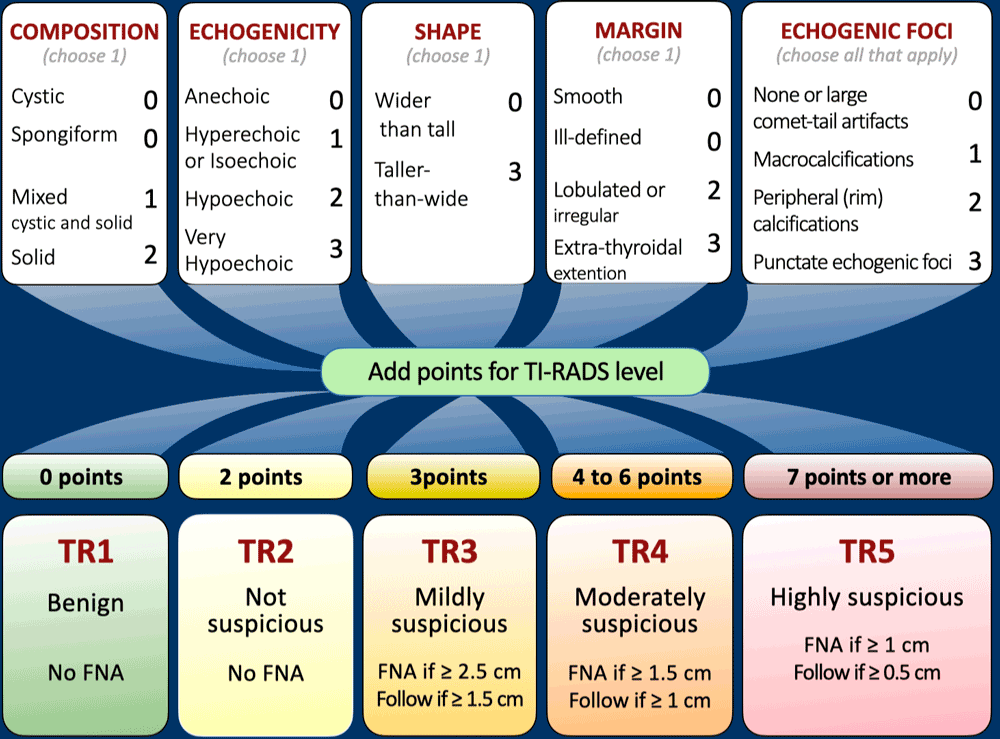
Tabla de clasificación de TIRADS
|
| Categoría |
Agujas |
Suspicion |
Riesgo de malignidad |
Pauta |
| TR1 |
0 |
Benigno |
0.3% |
Sin FNA |
| TR2 |
2 |
No sospechoso |
1.5% |
Sin FNA |
| TR3 |
3 |
Ligeramente sospechoso |
4.8% |
Si ≥2,5 cm: PAAF Si ≥1,5 cm: Seguimiento a los 1, 3 y 5 años
|
| TR4 |
4-6 |
Moderadamente sospechoso |
9.1% |
Si ≥1,5 cm: PAAF Si ≥1 cm: Seguimiento a los 1, 3 y 5 años
|
| TR5 |
7 o más |
Altamente sospechoso |
35% |
Si ≥1 cm: PAAF Si ≥0,5 cm, realizar seguimiento anual durante 5 años
|
FNA-Aspiración con aguja fina.
Riesgo de malignidad de tiroides según la puntuación TIRADS
- Los nódulos tiroideos TR 1 tienen un riesgo de malignidad del 0,3 %.
- Los nódulos tiroideos TR 2 tienen un riesgo de malignidad del 1,5 %.
- Los nódulos tiroideos TR 3 tienen un riesgo de malignidad del 4,8 %.
- Los nódulos tiroideos TR 4 tienen un riesgo de malignidad del 9,1 %.
- Los nódulos tiroideos TR 5 tienen un riesgo de malignidad del 35 %.
Advertencias sobre la calculadora de ecografía de tiroides
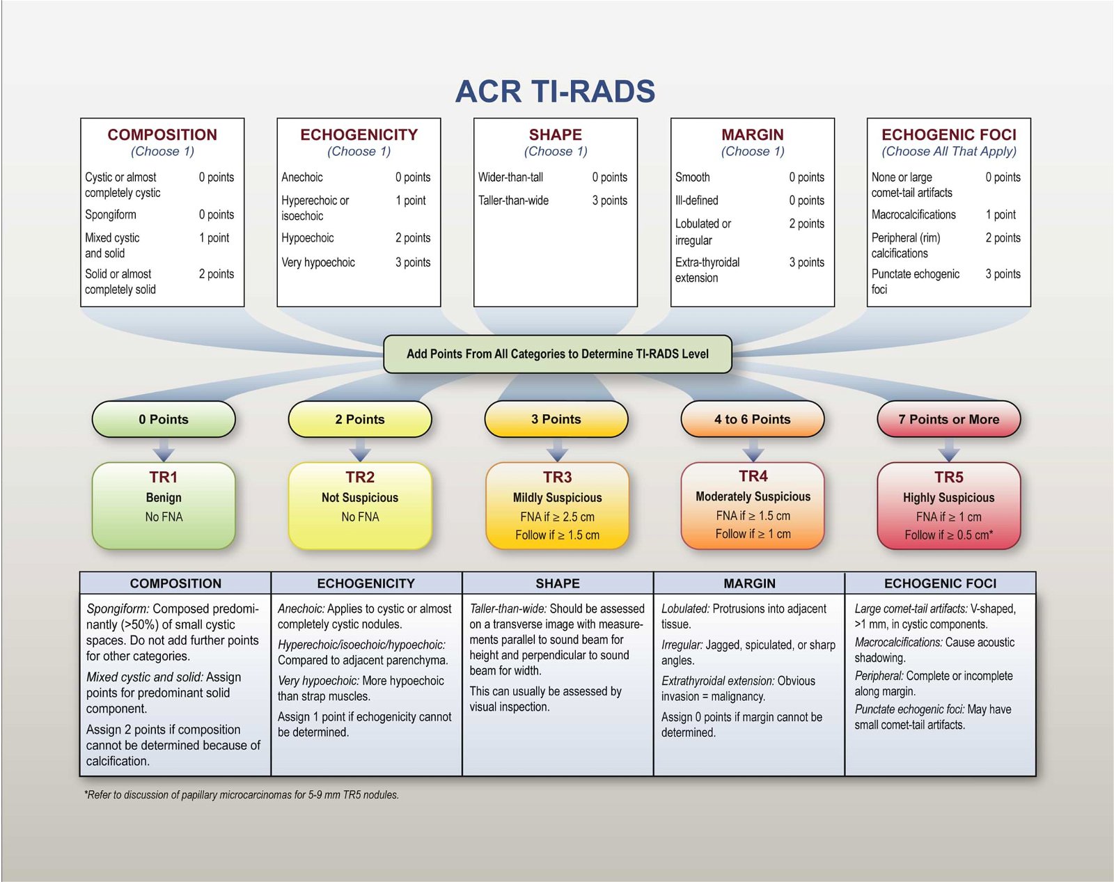
- Si las calcificaciones del borde ocultan completamente el nódulo, elija una composición “sólida” y una ecogenicidad “isoecoica”.
- Si no se puede determinar el margen, elija “margen mal definido”.
- Si no se puede determinar la ecogenicidad, elija “isoecoico”.
- Si no se puede determinar la composición, elija “sólido”.
- Los focos ecogénicos puntiformes y las características más altas que anchas tienen los puntos más altos, por lo que deben evaluarse con cuidado.
Puntos clave del artículo original de TIRADS:
Composición
Los nódulos quísticos o casi completamente quísticos son generalmente benignos, al igual que los nódulos espongiformes, compuestos predominantemente por pequeños espacios quísticos. Los componentes sólidos con características sospechosas pueden requerir una evaluación adicional para detectar malignidad.
Ecogenicidad
La evaluación de la ecogenicidad implica comparar la reflectividad de un nódulo con el tejido tiroideo adyacente, excepto en el caso de nódulos muy hipoecoicos, donde los músculos de la banda sirven como punto de referencia. Se presta especial atención a los nódulos anecoicos.
Forma
Una forma más alta que ancha, cuando se evalúa en el plano axial comparando las medidas de altura y ancho, puede ser altamente específica de malignidad.
Margen
Los márgenes mal definidos o irregulares, especialmente con protuberancias o lobulaciones, deben despertar sospechas. También debe considerarse la extensión más allá del borde tiroideo.
Focos ecogénicos
Los diferentes tipos de focos ecogénicos conllevan distintos niveles de sospecha. Los focos ecogénicos puntiformes dentro de componentes sólidos son particularmente preocupantes y deben evaluarse junto con otras características.
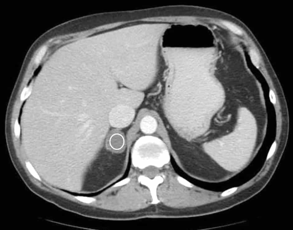
Microcarcinomas papilares de tiroides
Generalmente no se recomienda la biopsia de nódulos menores de 1 cm; sin embargo, se pueden hacer excepciones según circunstancias específicas. Las guías recomiendan no realizar biopsias rutinarias de estos nódulos pequeños, a menos que se realice una vigilancia activa, se realice una ablación o se considere una lobectomía para microcarcinomas papilares.
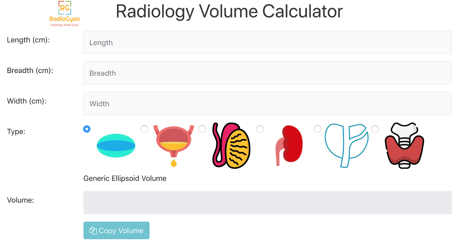
Medición y documentación
El dimensionamiento preciso y la documentación detallada de la ubicación de los nódulos son cruciales para el monitoreo y la comparación a lo largo del tiempo.
Definición de crecimiento
El agrandamiento significativo se define como un aumento del 20% en al menos dos dimensiones del nódulo con un aumento mínimo de 2 mm o un aumento del 50% o más en el volumen.
Momento de las ecografías de seguimiento
- No existe consenso en la literatura sobre el espaciamiento óptimo para las ecografías de seguimiento de los nódulos que no cumplen los criterios de tamaño de la PAAF.
- Por lo general no se recomiendan intervalos de exploración inferiores a un año, excepto en el caso de cánceres comprobados bajo vigilancia activa.
- Los intervalos de seguimiento deben basarse en el nivel ACR TI-RADS del nódulo; las lesiones más sospechosas requieren ecografías adicionales.
- Para las lesiones TR5, se recomiendan exploraciones anuales durante hasta 5 años.
- Para las lesiones TR4, se recomiendan exploraciones a los 1, 2, 3 y 5 años.
- Las lesiones TR3 pueden someterse a imágenes de seguimiento a los 1, 3 y 5 años.
- Las imágenes pueden suspenderse a los 5 años si no hay cambio de tamaño, indicativo de un comportamiento benigno.
- El tratamiento de los nódulos que aumentan de tamaño significativamente pero que permanecen por debajo del umbral de tamaño de la PAAF a los 5 años carece de orientación publicada, pero es probable que se requiera un seguimiento continuo.
- Si el nivel ACR TI-RADS de un nódulo aumenta durante el seguimiento, la siguiente ecografía debe programarse en 1 año, independientemente de su nivel inicial.
Número de nódulos a biopsiar
Se recomienda biopsiar los dos nódulos con mayor apariencia sospechosa para evitar procedimientos innecesarios y molestias al paciente.
Evaluación de los ganglios linfáticos cervicales
Las características específicas que indican metástasis deben motivar la realización de una PAAF de ganglios sospechosos junto con hasta dos nódulos que cumplan los criterios de biopsia según las pautas TI-RADS del ACR.
Preguntas frecuentes sobre TI-RADS
¿Qué significan las siglas TI-RADS?
TI-RADS son las siglas de Sistema de Informes y Datos de Imagen Tiroidea. Estandariza los informes ecográficos y el manejo de los nódulos tiroideos mediante un sistema basado en puntos del Colegio Americano de Radiología (ACR).
¿Cómo utilizo la calculadora TI-RADS?
1) Realizar una ecografía tiroidea completa. 2) Seleccionar las características de composición, ecogenicidad, forma, márgenes y focos ecogénicos. 3) Revisar la puntuación total y la categoría TI-RADS. 4) Seguir las recomendaciones de seguimiento o PAAF según el tamaño de la lesión.
¿Cuáles son las categorías TI-RADS?
TR1 Benigno, TR2 No sospechoso, TR3 Levemente sospechoso, TR4 Moderadamente sospechoso, TR5 Altamente sospechoso. La categoría se determina por la puntuación total asignada a las características ecográficas.
¿Qué características ecográficas se evalúan?
Cinco dominios: composición, ecogenicidad, forma (transversal), margen y focos ecogénicos. Cada dominio aporta puntos que, en conjunto, dan como resultado una categoría TI-RADS.
¿Cuándo se debe realizar una PAAF?
Umbrales típicos de tamaño según la clasificación ACR: TR3 considera FNA ≥2,5 cm; TR4 considera FNA ≥1,5 cm; TR5 recomienda FNA ≥1,0 cm. Aplique siempre el criterio clínico.
¿Qué son los intervalos de seguimiento?
Enfoque habitual: TR1–TR2 sin seguimiento rutinario; TR3: ecografía a los 12 meses aproximadamente; TR4: ecografía a los 6–12 meses; TR5: ecografía a los 3–6 meses si no se ha realizado biopsia. Modificar según el tamaño y el riesgo.
¿Qué tan preciso es TI-RADS?
El sistema ACR TI-RADS está validado y se utiliza ampliamente, mejorando la consistencia y reduciendo las biopsias innecesarias. Su precisión depende de la calidad de la imagen y de la correcta selección de características; no reemplaza el criterio clínico.
¿Qué características sugieren benignidad?
Composición espongiforme, nódulos anecoicos/quísticos, ecogenicidad hiperecoica/isoecoica, forma más ancha que alta, márgenes lisos y grandes artefactos de cola de cometa en los componentes quísticos.
¿Qué características aumentan las sospechas?
Ecogenicidad muy hipoecoica, composición sólida, forma más alta que ancha, márgenes irregulares/lobulados, focos ecogénicos puntiformes, calcificaciones periféricas/macrocalcificaciones y extensión extratiroidea.
¿Cuáles son las limitaciones de TI-RADS?
Sistema basado únicamente en ultrasonido; variabilidad interobservador; no incluye directamente factores de riesgo clínicos ni la tasa de crecimiento; menos fiable en enfermedades difusas. Utilizar en el contexto clínico.
¿Esta herramienta se basa en las directrices de la ACR?
Sí, la puntuación se ajusta a los documentos técnicos y al léxico de ACR TI-RADS. La herramienta es educativa y no cuenta con el respaldo oficial de ACR.
¿Cómo aplicar el sistema TI-RADS en el cáncer de tiroides multinodular?
Evalúe cada nódulo individualmente. Realice una PAAF a la vez para no más de dos nódulos con las puntuaciones más altas; realice el seguimiento de no más de cuatro nódulos con las puntuaciones más altas.
¿Influye el tamaño del nódulo en el tratamiento?
El tamaño no modifica la puntuación TI-RADS, pero sí determina los umbrales de manejo para la PAAF y el seguimiento. Los nódulos de mayor tamaño dentro de las categorías superiores requieren una intervención más temprana.
¿Quién desarrolló esta calculadora?
Esta calculadora TI-RADS fue desarrollada y es mantenida por el Dr. Amar Udare, MD, DNB, radiólogo certificado y profesor clínico asociado en diagnóstico por imágenes (radiología) en la Universidad de Calgary.
Explicación de la puntuación ecográfica de la tiroides
Para obtener una guía más detallada con ejemplos, consulte el artículo dedicado aquí:
TIRADS ACR: ¡Lo que los radiólogos necesitan saber!
Más recursos


Sobre el Autor
Dr. Amar Udare, MD, DNB
El Dr. Udare es licenciado en Medicina y tiene una licenciatura en Medicina, y su especialidad es la radiología. Es autor de numerosas publicaciones con revisión por pares, contribuyendo significativamente al campo de la medicina. Sus trabajos están disponibles en PubMed y Google Académico .
Además de sus logros académicos y profesionales, el Dr. Udare es un ávido lector y disfruta explorando los últimos avances en tecnología médica. Su compromiso de hacer accesible el conocimiento médico complejo a los pacientes y al público en general se alinea con nuestra misión en RadioGyan.com.
Para cualquier pregunta o aclaración adicional, no dude en comunicarse con el Dr. Udare a través del formulario de contacto .