


Imaging of Endometriosis (Case-based approach) | Radiology Board Review Case
Pathophysiology of endometriosis
- Functional endometrial glands OUTSIDE the uterus.
- Reproductive age group.
- Chronic pain and infertility.
- Potential mechanisms
- Types
- Superficial
- Ovarian
- Deep pelvic endometriosis
Ultrasound features of endometriosis
- Homogeneous, hypoechoic focal lesion.
- Can be unilocular or multilocular
- Low-level internal echoes.
- Echogenic wall foci – Best single predictor.
- No vascularity.
How to differentiate hemorrhagic cyst from endometriosis?
Features of hemorrhagic cyst:
- Acute history
- Evolve into more complex appearing cysts.
- Fibrin strands are thinner than septa seen in endometriomas
- Absent echogenic wall foci
- Resolves in 4-6 weeks.
MR imaging of endometriosis
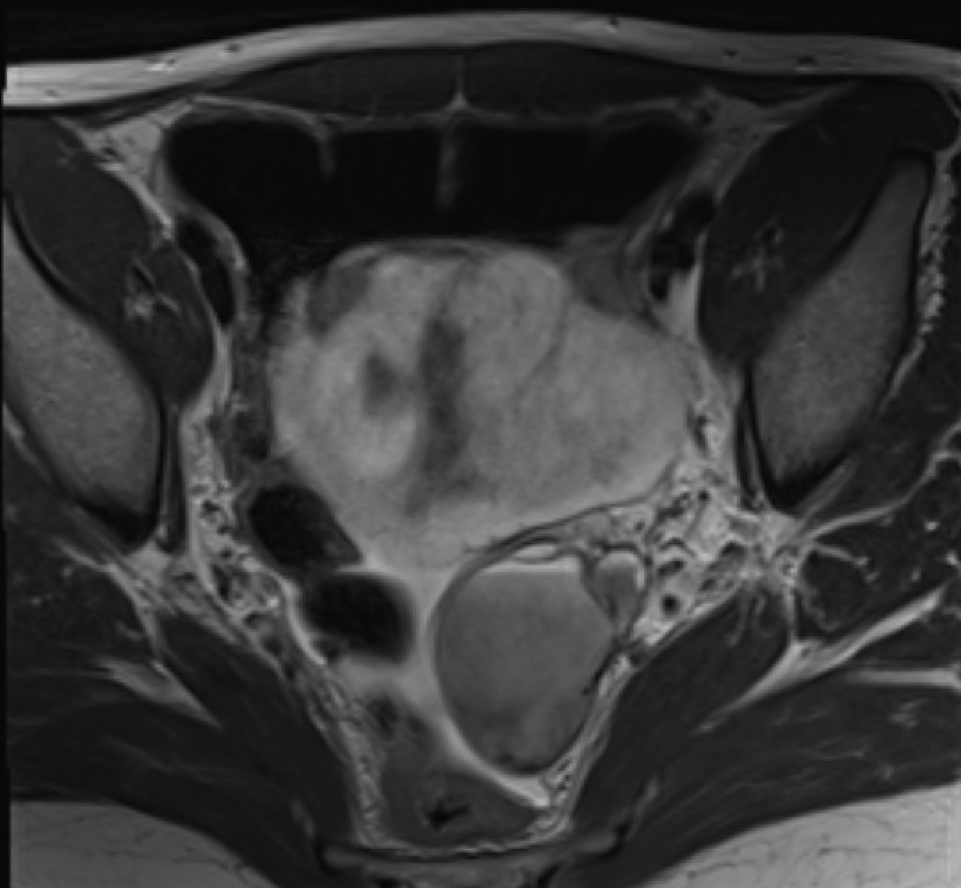
- Include T1 fat-saturated sequence.
- T2 shading: Cyst that is hyperintense on a T1-weighted image becomes hypointense on a T2-weighted image.
- Reflects the chronic nature of an endometrioma and helps differentiate it from other blood-containing lesions.
- Deep pelvic endometriosis
- Differentials: Hemorrhagic cyst, dermoid cyst, mucinous carcinoma.
- Watch the video for detailed PACS based case images.
MRI imaging pearls for Endometriosis
- Include T1 fat-sat sequence in MRI female pelvis.
- Multiple T1 hyperintense ovarian lesions = Endometriomas.
- Low Signal Intensity of Adnexal Masses on STIR MR Images Is Not Specific for Mature Cystic Teratoma and Does Not Exclude Endometrioma
- Benign Endometriomas Show Restricted Diffusion.
- Hematosalpinx Should Be Considered Specific for Pelvic Endometriosis
- Obstruction of Antegrade Menstrual Flow Increases the Risk for Endometriosis.
- Endometriomas Can Transform into Clear Cell or Endometrioid Epithelial Ovarian Carcinomas -Development of enhancing mural nodules.
- Solid Fibrotic Masses of Endometriosis Are Common and Easily Overlooked -Uterosacral Ligaments, rectovaginal pouch, bladder
- Solid Invasive Endometriosis of the Posterior Uterus Can Mimic Posterior Segmental Adenomyosis.
- Decidualized Endometriosis May Mimic Ovarian Malignancy in Pregnant Women – Vascular mural nodules
Reference and further reading
To attend live, join our Telegram group to get regular updates for these webinars:
More Radiology videos:


Improve Content And Help Your Colleagues!
Found an error in the post? Please let us know using our contact page and we will update it with due credits!
If you wish to contribute radiological images for this case (or any other article on the website), you can submit them here and you will be duly credited: Submit radiology case